4ZM9 | pdb_00004zm9

Crystal structure of circularly permuted human asparaginase-like protein 1

  • Classification: HYDROLASE
  • Organism(s): Homo sapiens
  • Expression System: Escherichia coli BL21(DE3)
  • Mutation(s): No 

  • Deposited: 2015-05-02 Released: 2016-02-17 
  • Deposition Author(s): Li, W.Z., Zhang, Y.
  • Funding Organization(s): National Institutes of Health/National Cancer Institute (NIH/NCI), Cancer Prevention and Research Institute of Texas (CPRIT)

Experimental Data Snapshot

  • Method: X-RAY DIFFRACTION
  • Resolution: 2.51 Å
  • R-Value Free: 
    0.233 (Depositor), 0.238 (DCC) 
  • R-Value Work: 
    0.204 (Depositor), 0.209 (DCC) 
  • R-Value Observed: 
    0.206 (Depositor) 

wwPDB Validation 3D Report Full Report

Validation slider image for 4ZM9

This is version 1.5 of the entry. See complete history

Literature

Intramolecular Cleavage of the hASRGL1 Homodimer Occurs in Two Stages.

Li, W.Irani, S.Crutchfield, A.Hodge, K.Matthews, W.Patel, P.Zhang, Y.J.Stone, E.

(2016) Biochemistry 55: 960-969

  • DOI: https://doi.org/10.1021/acs.biochem.5b01157
  • Primary Citation Related Structures: 
    4ZM9

  • PubMed Abstract: 

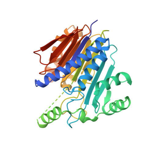
    The human asparaginase-like protein 1 (hASRGL1) is a member of the N-terminal nucleophile (Ntn) family that hydrolyzes l-asparagine and isoaspartyl-dipeptides. The nascent protein folds into an αβ-βα sandwich fold homodimer that cleaves its own peptide backbone at the G167-T168 bond, resulting in the active form of the enzyme. However, biophysical studies of hASRGL1 are difficult because of the curious fact that intramolecular cleavage of the G167-T168 peptide bond reaches only ≤50% completion. We capitalized upon our previous observation that intramolecular processing increases thermostability and developed a differential scanning fluorimetry assay that allowed direct detection of distinct processing intermediates for the first time. A kinetic analysis of these intermediates revealed that cleavage of one subunit of the hASRGL1 subunit drastically reduces the processing rate of the adjacent monomer, and a mutagenesis study showed that stabilization of the dimer interface plays a critical role in this process. We also report a comprehensive analysis of conserved active site residues and delineate their relative roles in autoprocessing and substrate hydrolysis. In addition to glycine, which was previously reported to selectively accelerate hASRGL1 cleavage, we identified several novel small molecule activators that also promote intramolecular processing. The structure-activity analysis supports the hypothesis that multiple negatively charged small molecules interact within the active site of hASRGL1 to act as a base in promoting cleavage. Overall, our investigation provides a mechanistic understanding of the maturation process of this Ntn hydrolase family member.


  • Organizational Affiliation
    • Department of Molecular Biosciences, ‡Department of Chemical Engineering, and §Institute of Cellular and Molecular Biology, University of Texas , Austin, Texas 78712, United States.

Macromolecule Content 

  • Total Structure Weight: 137 kDa 
  • Atom Count: 8,838 
  • Modeled Residue Count: 1,184 
  • Deposited Residue Count: 1,308 
  • Unique protein chains: 1

Macromolecules

Find similar proteins by:|  3D Structure
Entity ID: 1
MoleculeChains  Sequence LengthOrganismDetailsImage
Isoaspartyl peptidase/L-asparaginase
A, B, C, D
327Homo sapiensMutation(s): 0 
Gene Names: ASRGL1ALPCRASH
EC: 3.4.19.5 (PDB Primary Data), 3.5.1.1 (PDB Primary Data)
UniProt & NIH Common Fund Data Resources
Find proteins for Q7L266 (Homo sapiens)
Explore Q7L266 
Go to UniProtKB:  Q7L266
PHAROS:  Q7L266
GTEx:  ENSG00000162174 
Entity Groups
Sequence Clusters30% Identity50% Identity70% Identity90% Identity95% Identity100% Identity
UniProt GroupQ7L266
Sequence Annotations
Expand
Reference Sequence

Small Molecules

Ligands 5 Unique
IDChains Name / Formula / InChI Key2D Diagram3D Interactions
FLC

Query on FLC


Download:Ideal Coordinates CCD File
BA [auth D],
O [auth B]
CITRATE ANION
C6 H5 O7
KRKNYBCHXYNGOX-UHFFFAOYSA-K
DTT

Query on DTT


Download:Ideal Coordinates CCD File
K [auth A],
W [auth C]
2,3-DIHYDROXY-1,4-DITHIOBUTANE
C4 H10 O2 S2
VHJLVAABSRFDPM-IMJSIDKUSA-N
BME

Query on BME


Download:Ideal Coordinates CCD File
CA [auth D]
G [auth A]
H [auth A]
I [auth A]
J [auth A]
CA [auth D],
G [auth A],
H [auth A],
I [auth A],
J [auth A],
P [auth B],
S [auth C],
T [auth C],
U [auth C],
V [auth C]
BETA-MERCAPTOETHANOL
C2 H6 O S
DGVVWUTYPXICAM-UHFFFAOYSA-N
GLY

Query on GLY


Download:Ideal Coordinates CCD File
AA [auth D],
F [auth A],
N [auth B],
R [auth C]
GLYCINE
C2 H5 N O2
DHMQDGOQFOQNFH-UHFFFAOYSA-N
NA

Query on NA


Download:Ideal Coordinates CCD File
E [auth A]
L [auth A]
M [auth B]
Q [auth C]
X [auth C]
E [auth A],
L [auth A],
M [auth B],
Q [auth C],
X [auth C],
Y [auth C],
Z [auth D]
SODIUM ION
Na
FKNQFGJONOIPTF-UHFFFAOYSA-N

Experimental Data & Validation

Experimental Data

  • Method: X-RAY DIFFRACTION
  • Resolution: 2.51 Å
  • R-Value Free:  0.233 (Depositor), 0.238 (DCC) 
  • R-Value Work:  0.204 (Depositor), 0.209 (DCC) 
  • R-Value Observed: 0.206 (Depositor) 
Space Group: P 21 21 21
Unit Cell:
Length ( Å )Angle ( ˚ )
a = 104.308α = 90
b = 111.107β = 90
c = 122.517γ = 90
Software Package:
Software NamePurpose
REFMACrefinement
HKL-2000data reduction
HKL-2000data scaling
PHENIXphasing

Structure Validation

View Full Validation Report


Entry History 

& Funding Information

Deposition Data


Funding OrganizationLocationGrant Number
National Institutes of Health/National Cancer Institute (NIH/NCI)United StatesR01 CA154754
National Institutes of Health/National Cancer Institute (NIH/NCI)United StatesCA139059
Cancer Prevention and Research Institute of Texas (CPRIT)United StatesRP120314

Revision History  (Full details and data files)

  • Version 1.0: 2016-02-17
    Type: Initial release
  • Version 1.1: 2016-02-24
    Changes: Database references
  • Version 1.2: 2017-09-20
    Changes: Author supporting evidence, Database references, Derived calculations
  • Version 1.3: 2019-04-24
    Changes: Author supporting evidence, Data collection
  • Version 1.4: 2019-12-04
    Changes: Author supporting evidence
  • Version 1.5: 2024-03-06
    Changes: Data collection, Database references, Derived calculations, Refinement description