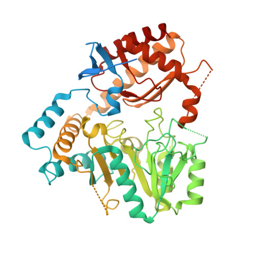
4ZM3

Crystal structure of PLP-Dependent 3-Aminobenzoate Synthase PctV wild-type


Experimental Data Snapshot

  • Method: X-RAY DIFFRACTION
  • Resolution: 2.27 Å
  • R-Value Free: 0.222 
  • R-Value Work: 0.180 
  • R-Value Observed: 0.182 

wwPDB Validation   3D Report Full Report


Ligand Structure Quality Assessment 


This is version 1.3 of the entry. See complete history


Literature

Mechanism-Based Trapping of the Quinonoid Intermediate by Using the K276R Mutant of PLP-Dependent 3-Aminobenzoate Synthase PctV in the Biosynthesis of Pactamycin.

Hirayama, A.Miyanaga, A.Kudo, F.Eguchi, T.

(2015) Chembiochem 16: 2484-2490

  • DOI: https://doi.org/10.1002/cbic.201500426
  • Primary Citation of Related Structures:  
    4ZM3, 4ZM4

  • PubMed Abstract: 

    Mutational analysis of the pyridoxal 5'-phosphate (PLP)-dependent enzyme PctV was carried out to elucidate the multi-step reaction mechanism for the formation of 3-aminobenzoate (3-ABA) from 3-dehydroshikimate (3-DSA). Introduction of mutation K276R led to the accumulation of a quinonoid intermediate with an absorption maximum at 580 nm after the reaction of pyridoxamine 5'-phosphate (PMP) with 3-DSA. The chemical structure of this intermediate was supported by X-ray crystallographic analysis of the complex formed between the K276R mutant and the quinonoid intermediate. These results clearly show that a quinonoid intermediate is involved in the formation of 3-ABA. They also indicate that Lys276 (in the active site of PctV) plays multiple roles, including acid/base catalysis during the dehydration reaction of the quinonoid intermediate.


  • Organizational Affiliation

    Department of Chemistry and Materials Science, Tokyo Institute of Technology, 2-12-1 O-okayama, Meguro-ku, Tokyo, 152-8551, Japan.


Macromolecules
Find similar proteins by:  (by identity cutoff)  |  3D Structure
Entity ID: 1
MoleculeChains Sequence LengthOrganismDetailsImage
Aminotransferase
A, B, C, D
464Streptomyces pactumMutation(s): 0 
Gene Names: pctVptmT
UniProt
Find proteins for A8R0K5 (Streptomyces pactum)
Explore A8R0K5 
Go to UniProtKB:  A8R0K5
Entity Groups  
Sequence Clusters30% Identity50% Identity70% Identity90% Identity95% Identity100% Identity
UniProt GroupA8R0K5
Sequence Annotations
Expand
  • Reference Sequence
Small Molecules
Ligands 3 Unique
IDChains Name / Formula / InChI Key2D Diagram3D Interactions
PLP
Query on PLP

Download Ideal Coordinates CCD File 
E [auth A],
F [auth B],
G [auth C],
I [auth D]
PYRIDOXAL-5'-PHOSPHATE
C8 H10 N O6 P
NGVDGCNFYWLIFO-UHFFFAOYSA-N
PGE
Query on PGE

Download Ideal Coordinates CCD File 
H [auth C]TRIETHYLENE GLYCOL
C6 H14 O4
ZIBGPFATKBEMQZ-UHFFFAOYSA-N
PEG
Query on PEG

Download Ideal Coordinates CCD File 
J [auth D],
K [auth D],
L [auth D]
DI(HYDROXYETHYL)ETHER
C4 H10 O3
MTHSVFCYNBDYFN-UHFFFAOYSA-N
Experimental Data & Validation

Experimental Data

  • Method: X-RAY DIFFRACTION
  • Resolution: 2.27 Å
  • R-Value Free: 0.222 
  • R-Value Work: 0.180 
  • R-Value Observed: 0.182 
  • Space Group: P 1 21 1
Unit Cell:
Length ( Å )Angle ( ˚ )
a = 58.99α = 90
b = 170.83β = 98.33
c = 74.57γ = 90
Software Package:
Software NamePurpose
REFMACrefinement
MOSFLMdata reduction
SCALAdata scaling
MOLREPphasing
Cootmodel building
ARPmodel building

Structure Validation

View Full Validation Report


Ligand Structure Quality Assessment 


Entry History & Funding Information

Deposition Data


Funding OrganizationLocationGrant Number
Ministry of Education, Culture, Sports, Science and TechnologyJapan22108003

Revision History  (Full details and data files)

  • Version 1.0: 2015-10-14
    Type: Initial release
  • Version 1.1: 2016-01-27
    Changes: Database references
  • Version 1.2: 2020-02-19
    Changes: Data collection, Derived calculations
  • Version 1.3: 2023-11-08
    Changes: Data collection, Database references, Refinement description