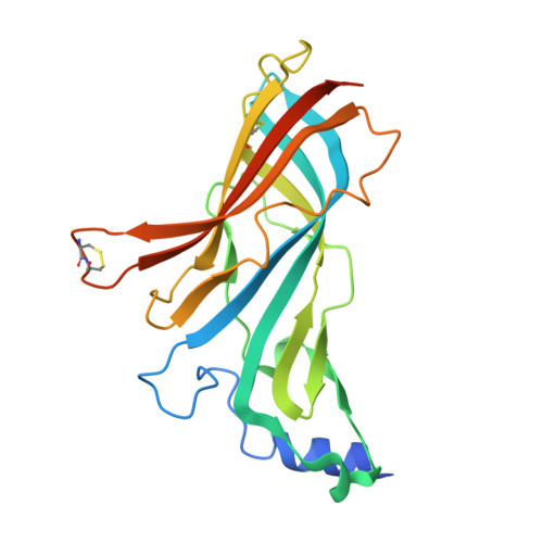
4ZJS

Crystal structure of a chimeric acetylcholine binding protein from Aplysia Californica (Ac-AChBP) containing the main immunogenic region (MIR) from the human alpha 1 subunit of the muscle nicotinic acetylcholine receptor in complex with anatoxin-A.


Experimental Data Snapshot

  • Method: X-RAY DIFFRACTION
  • Resolution: 2.23 Å
  • R-Value Free: 0.228 
  • R-Value Work: 0.197 
  • R-Value Observed: 0.197 

Starting Models: experimental
View more details

wwPDB Validation   3D Report Full Report


Ligand Structure Quality Assessment 


This is version 1.1 of the entry. See complete history


Literature

Main immunogenic region structure promotes binding of conformation-dependent myasthenia gravis autoantibodies, nicotinic acetylcholine receptor conformation maturation, and agonist sensitivity.

Luo, J.Taylor, P.Losen, M.de Baets, M.H.Shelton, G.D.Lindstrom, J.

(2009) J Neurosci 29: 13898-13908

  • DOI: https://doi.org/10.1523/JNEUROSCI.2833-09.2009
  • Primary Citation of Related Structures:  
    4ZJS

  • PubMed Abstract: 

    The main immunogenic region (MIR) is a conformation-dependent region at the extracellular apex of alpha1 subunits of muscle nicotinic acetylcholine receptor (AChR) that is the target of half or more of the autoantibodies to muscle AChRs in human myasthenia gravis and rat experimental autoimmune myasthenia gravis. By making chimeras of human alpha1 subunits with alpha7 subunits, both MIR epitopes recognized by rat mAbs and by the patient-derived human mAb 637 to the MIR were determined to consist of two discontiguous sequences, which are adjacent only in the native conformation. The MIR, including loop alpha1 67-76 in combination with the N-terminal alpha helix alpha1 1-14, conferred high-affinity binding for most rat mAbs to the MIR. However, an additional sequence corresponding to alpha1 15-32 was required for high-affinity binding of human mAb 637. A water soluble chimera of Aplysia acetylcholine binding protein with the same alpha1 MIR sequences substituted was recognized by a majority of human, feline, and canine myasthenia gravis sera. The presence of the alpha1 MIR sequences in alpha1/alpha7 chimeras greatly promoted AChR expression and significantly altered the sensitivity to activation. This reveals a structural and functional, as well as antigenic, significance of the MIR.


  • Organizational Affiliation

    Department of Neuroscience, University of Pennsylvania Medical School, Philadelphia, PA 19104-6074, USA.


Macromolecules
Find similar proteins by:  (by identity cutoff)  |  3D Structure
Entity ID: 1
MoleculeChains Sequence LengthOrganismDetailsImage
Acetylcholine receptor subunit alpha,Soluble acetylcholine receptor,Acetylcholine receptor subunit alpha,Soluble acetylcholine receptor
A, B, C, D, E
230Homo sapiensAplysia californicaMutation(s): 0 
Gene Names: CHRNA1ACHRACHNRA
UniProt & NIH Common Fund Data Resources
Find proteins for P02708 (Homo sapiens)
Explore P02708 
Go to UniProtKB:  P02708
PHAROS:  P02708
GTEx:  ENSG00000138435 
Find proteins for Q8WSF8 (Aplysia californica)
Explore Q8WSF8 
Go to UniProtKB:  Q8WSF8
Entity Groups  
Sequence Clusters30% Identity50% Identity70% Identity90% Identity95% Identity100% Identity
UniProt GroupsQ8WSF8P02708
Sequence Annotations
Expand
  • Reference Sequence
Experimental Data & Validation

Experimental Data

  • Method: X-RAY DIFFRACTION
  • Resolution: 2.23 Å
  • R-Value Free: 0.228 
  • R-Value Work: 0.197 
  • R-Value Observed: 0.197 
  • Space Group: I 2 3
Unit Cell:
Length ( Å )Angle ( ˚ )
a = 207.604α = 90
b = 207.604β = 90
c = 207.604γ = 90
Software Package:
Software NamePurpose
PHENIXrefinement
HKL-2000data scaling
PDB_EXTRACTdata extraction
HKL-2000data reduction
PHASERphasing

Structure Validation

View Full Validation Report


Ligand Structure Quality Assessment 


Entry History 

Deposition Data

Revision History  (Full details and data files)

  • Version 1.0: 2015-05-13
    Type: Initial release
  • Version 1.1: 2023-09-27
    Changes: Advisory, Data collection, Database references, Derived calculations, Refinement description, Source and taxonomy