4ZIV | pdb_00004ziv

Crystal structure of AcrB triple mutant in P21 space group


Experimental Data Snapshot

  • Method: X-RAY DIFFRACTION
  • Resolution: 3.16 Å
  • R-Value Free: 
    0.335 (Depositor), 0.364 (DCC) 
  • R-Value Work: 
    0.270 (Depositor) 
  • R-Value Observed: 
    0.273 (Depositor) 

Starting Model: experimental
View more details

wwPDB Validation 3D Report Full Report

Validation slider image for 4ZIV

Ligand Structure Quality Assessment 


This is version 1.2 of the entry. See complete history

Literature

Structures of Gate Loop Variants of the AcrB Drug Efflux Pump Bound by Erythromycin Substrate.

Ababou, A.Koronakis, V.

(2016) PLoS One 11: e0159154-e0159154

  • DOI: https://doi.org/10.1371/journal.pone.0159154
  • Primary Citation Related Structures: 
    4ZIT, 4ZIV, 4ZIW, 4ZJL, 4ZJO, 4ZJQ

  • PubMed Abstract: 

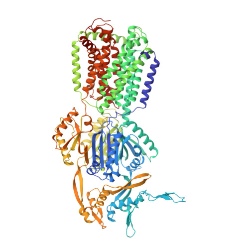
    Gram-negative bacteria such as E. coli use tripartite efflux pumps such as AcrAB-TolC to expel antibiotics and noxious compounds. A key feature of the inner membrane transporter component, AcrB, is a short stretch of residues known as the gate/switch loop that divides the proximal and distal substrate binding pockets. Amino acid substitutions of the gate loop are known to decrease antibiotic resistance conferred by AcrB. Here we present two new AcrB gate loop variants, the first stripped of its bulky side chains, and a second in which the gate loop is removed entirely. By determining the crystal structures of the variant AcrB proteins in the presence and absence of erythromycin and assessing their ability to confer erythromycin tolerance, we demonstrate that the gate loop is important for AcrB export activity but is not required for erythromycin binding.


  • Organizational Affiliation
    • Department of Pathology, University of Cambridge, Tennis Court Road, Cambridge, United Kingdom.

Macromolecule Content 

  • Total Structure Weight: 683.8 kDa 
  • Atom Count: 47,736 
  • Modeled Residue Count: 6,262 
  • Deposited Residue Count: 6,294 
  • Unique protein chains: 1

Macromolecules

Find similar proteins by:|  3D Structure
Entity ID: 1
MoleculeChains  Sequence LengthOrganismDetailsImage
Multidrug efflux pump subunit AcrB
A, B, C, D, E
A, B, C, D, E, F
1,049Escherichia coli K-12Mutation(s): 3 
Gene Names: acrBacrEb0462JW0451
Membrane Entity: Yes 
UniProt
Find proteins for P31224 (Escherichia coli (strain K12))
Explore P31224 
Go to UniProtKB:  P31224
Entity Groups
Sequence Clusters30% Identity50% Identity70% Identity90% Identity95% Identity100% Identity
UniProt GroupP31224
Sequence Annotations
Expand
Reference Sequence

Small Molecules

Ligands 2 Unique
IDChains Name / Formula / InChI Key2D Diagram3D Interactions
LMT

Query on LMT


Download:Ideal Coordinates CCD File
G [auth A]
I [auth B]
J [auth C]
L [auth D]
M [auth E]
G [auth A],
I [auth B],
J [auth C],
L [auth D],
M [auth E],
O [auth F]
DODECYL-BETA-D-MALTOSIDE
C24 H46 O11
NLEBIOOXCVAHBD-QKMCSOCLSA-N
NI

Query on NI


Download:Ideal Coordinates CCD File
H [auth A],
K [auth C],
N [auth E]
NICKEL (II) ION
Ni
VEQPNABPJHWNSG-UHFFFAOYSA-N

Experimental Data & Validation

Experimental Data

  • Method: X-RAY DIFFRACTION
  • Resolution: 3.16 Å
  • R-Value Free:  0.335 (Depositor), 0.364 (DCC) 
  • R-Value Work:  0.270 (Depositor) 
  • R-Value Observed: 0.273 (Depositor) 
Space Group: P 1 21 1
Unit Cell:
Length ( Å )Angle ( ˚ )
a = 152.278α = 90
b = 157.491β = 92.74
c = 219.155γ = 90
Software Package:
Software NamePurpose
PHENIXrefinement
MOSFLMdata reduction
SCALAdata scaling
PHASERphasing

Structure Validation

View Full Validation Report


Ligand Structure Quality Assessment 


Entry History 

& Funding Information

Deposition Data


Funding OrganizationLocationGrant Number
Medical Research Council (United Kingdom)United Kingdom--

Revision History  (Full details and data files)

  • Version 1.0: 2016-07-27
    Type: Initial release
  • Version 1.1: 2017-09-13
    Changes: Author supporting evidence
  • Version 1.2: 2024-01-10
    Changes: Data collection, Database references, Derived calculations, Refinement description