4ZET

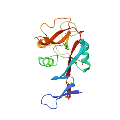
Blood dendritic cell antigen 2 (BDCA-2) complexed with GalGlcNAcMan


Experimental Data Snapshot

  • Method: X-RAY DIFFRACTION
  • Resolution: 2.90 Å
  • R-Value Free: 0.263 
  • R-Value Work: 0.178 
  • R-Value Observed: 0.182 

wwPDB Validation   3D Report Full Report


This is version 2.0 of the entry. See complete history


Literature

A Novel Mechanism for Binding of Galactose-terminated Glycans by the C-type Carbohydrate Recognition Domain in Blood Dendritic Cell Antigen 2.

Jegouzo, S.A.Feinberg, H.Dungarwalla, T.Drickamer, K.Weis, W.I.Taylor, M.E.

(2015) J Biol Chem 290: 16759-16771

  • DOI: https://doi.org/10.1074/jbc.M115.660613
  • Primary Citation of Related Structures:  
    4ZES, 4ZET

  • PubMed Abstract: 

    Blood dendritic cell antigen 2 (BDCA-2; also designated CLEC4C or CD303) is uniquely expressed on plasmacytoid dendritic cells. Stimulation of BDCA-2 with antibodies leads to an anti-inflammatory response in these cells, but the natural ligands for the receptor are not known. The C-type carbohydrate recognition domain in the extracellular portion of BDCA-2 contains a signature motif typical of C-type animal lectins that bind mannose, glucose, or GlcNAc, yet it has been reported that BDCA-2 binds selectively to galactose-terminated, biantennary N-linked glycans. A combination of glycan array analysis and binding competition studies with monosaccharides and natural and synthetic oligosaccharides have been used to define the binding epitope for BDCA-2 as the trisaccharide Galβ1-3/4GlcNAcβ1-2Man. X-ray crystallography and mutagenesis studies show that mannose is ligated to the conserved Ca(2+) in the primary binding site that is characteristic of C-type carbohydrate recognition domains, and the GlcNAc and galactose residues make additional interactions in a wide, shallow groove adjacent to the primary binding site. As predicted from these studies, BDCA-2 binds to IgG, which bears galactose-terminated glycans that are not commonly found attached to other serum glycoproteins. Thus, BDCA-2 has the potential to serve as a previously unrecognized immunoglobulin Fc receptor.


  • Organizational Affiliation

    the Department of Life Sciences, Imperial College, London SW7 2AZ, United Kingdom.


Macromolecules
Find similar proteins by:  (by identity cutoff)  |  3D Structure
Entity ID: 1
MoleculeChains Sequence LengthOrganismDetailsImage
C-type lectin domain family 4 member C
A, B
147Homo sapiensMutation(s): 1 
Gene Names: CLEC4CBDCA2CLECSF11CLECSF7DLECHECLUNQ9361/PRO34150
UniProt & NIH Common Fund Data Resources
Find proteins for Q8WTT0 (Homo sapiens)
Explore Q8WTT0 
Go to UniProtKB:  Q8WTT0
PHAROS:  Q8WTT0
GTEx:  ENSG00000198178 
Entity Groups  
Sequence Clusters30% Identity50% Identity70% Identity90% Identity95% Identity100% Identity
UniProt GroupQ8WTT0
Sequence Annotations
Expand
  • Reference Sequence
Oligosaccharides

Help

Entity ID: 2
MoleculeChains Length2D Diagram Glycosylation3D Interactions
beta-D-galactopyranose-(1-4)-2-acetamido-2-deoxy-beta-D-glucopyranose-(1-2)-alpha-D-mannopyranose
C, D
3N/A
Glycosylation Resources
GlyTouCan:  G28052FT
GlyCosmos:  G28052FT
GlyGen:  G28052FT
Experimental Data & Validation

Experimental Data

  • Method: X-RAY DIFFRACTION
  • Resolution: 2.90 Å
  • R-Value Free: 0.263 
  • R-Value Work: 0.178 
  • R-Value Observed: 0.182 
  • Space Group: P 2 21 21
Unit Cell:
Length ( Å )Angle ( ˚ )
a = 37.559α = 90
b = 67.645β = 90
c = 118.142γ = 90
Software Package:
Software NamePurpose
PHENIXrefinement
XDSdata reduction
Aimlessdata scaling
PHENIXphasing

Structure Validation

View Full Validation Report


Entry History & Funding Information

Deposition Data


Funding OrganizationLocationGrant Number
Wellcome TrustUnited Kingdom093599
National Institutes of Health/National Institute of General Medical Sciences (NIH/NIGMS)United StatesP41GM103393
National Institutes of Health/National Center for Research Resources (NIH/NCRR)United StatesP41RR001209
Department of Energy (DOE, United States)United StatesDE-AC02-06CH11357

Revision History  (Full details and data files)

  • Version 1.0: 2015-05-27
    Type: Initial release
  • Version 1.1: 2015-06-03
    Changes: Database references
  • Version 1.2: 2015-07-15
    Changes: Database references
  • Version 1.3: 2017-08-23
    Changes: Data collection, Database references, Derived calculations, Refinement description, Source and taxonomy
  • Version 1.4: 2017-09-06
    Changes: Author supporting evidence
  • Version 1.5: 2019-12-04
    Changes: Author supporting evidence
  • Version 2.0: 2020-07-29
    Type: Remediation
    Reason: Carbohydrate remediation
    Changes: Atomic model, Data collection, Derived calculations, Structure summary