


Crystal structure of TTK kinase domain in complex with a pyrazolopyrimidine inhibitor.
Qiu, W., Plotnikova, O., Feher, M., Awrey, D.E., Battaile, K., Chirgadze, N.Y.To be published.
Experimental Data Snapshot
Entity ID: 1 | |||||
|---|---|---|---|---|---|
| Molecule | Chains | Sequence Length | Organism | Details | Image |
| Dual specificity protein kinase TTK | 281 | Homo sapiens | Mutation(s): 0 Gene Names: TTK, MPS1, MPS1L1 EC: 2.7.12.1 | 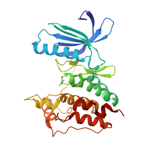 | |
UniProt & NIH Common Fund Data Resources | |||||
PHAROS: P33981 GTEx: ENSG00000112742 | |||||
Entity Groups | |||||
| Sequence Clusters | 30% Identity50% Identity70% Identity90% Identity95% Identity100% Identity | ||||
| UniProt Group | P33981 | ||||
Sequence AnnotationsExpand | |||||
Reference Sequence | |||||
| Ligands 4 Unique | |||||
|---|---|---|---|---|---|
| ID | Chains | Name / Formula / InChI Key | 2D Diagram | 3D Interactions | |
| 052 Download:Ideal Coordinates CCD File | J [auth A] | N-cyclopropyl-2-methyl-4-(7-{[2-(morpholin-4-yl)ethyl]amino}-5-phenoxypyrazolo[1,5-a]pyrimidin-3-yl)benzamide C29 H32 N6 O3 AKGWVDBXMHMMSD-UHFFFAOYSA-N |  | ||
| PEG Download:Ideal Coordinates CCD File | C [auth A] | DI(HYDROXYETHYL)ETHER C4 H10 O3 MTHSVFCYNBDYFN-UHFFFAOYSA-N |  | ||
| GOL Download:Ideal Coordinates CCD File | B [auth A] | GLYCEROL C3 H8 O3 PEDCQBHIVMGVHV-UHFFFAOYSA-N |  | ||
| EDO Download:Ideal Coordinates CCD File | D [auth A] E [auth A] F [auth A] G [auth A] H [auth A] | 1,2-ETHANEDIOL C2 H6 O2 LYCAIKOWRPUZTN-UHFFFAOYSA-N |  | ||
| Length ( Å ) | Angle ( ˚ ) |
|---|---|
| a = 70.311 | α = 90 |
| b = 106.693 | β = 90 |
| c = 111.86 | γ = 90 |
| Software Name | Purpose |
|---|---|
| REFMAC | refinement |
| XDS | data reduction |
| XDS | data scaling |


