Thiophenecarboxamide Derivatives Activated by EthA Kill Mycobacterium tuberculosis by Inhibiting the CTP Synthetase PyrG.
Mori, G., Chiarelli, L.R., Esposito, M., Makarov, V., Bellinzoni, M., Hartkoorn, R.C., Degiacomi, G., Boldrin, F., Ekins, S., de Jesus Lopes Ribeiro, A.L., Marino, L.B., Centarova, I., Svetlikova, Z., Blasko, J., Kazakova, E., Lepioshkin, A., Barilone, N., Zanoni, G., Porta, A., Fondi, M., Fani, R., Baulard, A.R., Mikusova, K., Alzari, P.M., Manganelli, R., de Carvalho, L.P., Riccardi, G., Cole, S.T., Pasca, M.R.(2015) Chem Biol 22: 917-927
- PubMed: 26097035 Search on PubMedSearch on PubMed Central
- DOI: https://doi.org/10.1016/j.chembiol.2015.05.016
- Primary Citation Related Structures:
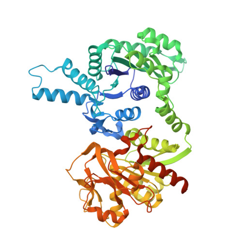
4ZDI, 4ZDJ, 4ZDK - PubMed Abstract:
To combat the emergence of drug-resistant strains of Mycobacterium tuberculosis, new antitubercular agents and novel drug targets are needed. Phenotypic screening of a library of 594 hit compounds uncovered two leads that were active against M. tuberculosis in its replicating, non-replicating, and intracellular states: compounds 7947882 (5-methyl-N-(4-nitrophenyl)thiophene-2-carboxamide) and 7904688 (3-phenyl-N-[(4-piperidin-1-ylphenyl)carbamothioyl]propanamide). Mutants resistant to both compounds harbored mutations in ethA (rv3854c), the gene encoding the monooxygenase EthA, and/or in pyrG (rv1699) coding for the CTP synthetase, PyrG. Biochemical investigations demonstrated that EthA is responsible for the activation of the compounds, and by mass spectrometry we identified the active metabolite of 7947882, which directly inhibits PyrG activity. Metabolomic studies revealed that pharmacological inhibition of PyrG strongly perturbs DNA and RNA biosynthesis, and other metabolic processes requiring nucleotides. Finally, the crystal structure of PyrG was solved, paving the way for rational drug design with this newly validated drug target.
- Department of Biology and Biotechnology "Lazzaro Spallanzani", University of Pavia, 27100 Pavia, Italy.
Organizational Affiliation: