4ZCM | pdb_00004zcm

Crystal Structure of Escherichia coli GTPase BipA/TypA Complexed with ppGpp


Experimental Data Snapshot

  • Method: X-RAY DIFFRACTION
  • Resolution: 3.31 Å
  • R-Value Free: 
    0.291 (Depositor), 0.297 (DCC) 
  • R-Value Work: 
    0.233 (Depositor), 0.240 (DCC) 
  • R-Value Observed: 
    0.235 (Depositor) 

Starting Model: experimental
View more details

wwPDB Validation 3D Report Full Report

Validation slider image for 4ZCM

Ligand Structure Quality Assessment 


This is version 1.3 of the entry. See complete history

Literature

Structural and Functional Analysis of BipA, a Regulator of Virulence in Enteropathogenic Escherichia coli.

Fan, H.Hahm, J.Diggs, S.Perry, J.J.Blaha, G.

(2015) J Biological Chem 290: 20856-20864

  • DOI: https://doi.org/10.1074/jbc.M115.659136
  • Primary Citation Related Structures: 
    4ZCI, 4ZCK, 4ZCL, 4ZCM

  • PubMed Abstract: 

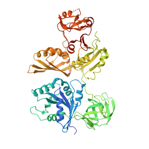
    The translational GTPase BipA regulates the expression of virulence and pathogenicity factors in several eubacteria. BipA-dependent expression of virulence factors occurs under starvation conditions, such as encountered during infection of a host. Under these conditions, BipA associates with the small ribosomal subunit. BipA also has a second function to promote the efficiency of late steps in biogenesis of large ribosomal subunits at low temperatures, presumably while bound to the ribosome. During starvation, the cellular concentration of stress alarmone guanosine-3', 5'-bis pyrophosphate (ppGpp) is increased. This increase allows ppGpp to bind to BipA and switch its binding specificity from ribosomes to small ribosomal subunits. A conformational change of BipA upon ppGpp binding could explain the ppGpp regulation of the binding specificity of BipA. Here, we present the structures of the full-length BipA from Escherichia coli in apo, GDP-, and ppGpp-bound forms. The crystal structure and small-angle x-ray scattering data of the protein with bound nucleotides, together with a thermodynamic analysis of the binding of GDP and of ppGpp to BipA, indicate that the ppGpp-bound form of BipA adopts the structure of the GDP form. This suggests furthermore, that the switch in binding preference only occurs when both ppGpp and the small ribosomal subunit are present. This molecular mechanism would allow BipA to interact with both the ribosome and the small ribosomal subunit during stress response.


  • Organizational Affiliation
    • Department of Biochemistry, University of California, Riverside, California 92521.

Macromolecule Content 

  • Total Structure Weight: 143.88 kDa 
  • Atom Count: 8,579 
  • Modeled Residue Count: 1,122 
  • Deposited Residue Count: 1,282 
  • Unique protein chains: 1

Macromolecules

Find similar proteins by:|  3D Structure
Entity ID: 1
MoleculeChains  Sequence LengthOrganismDetailsImage
GTP-binding protein TypA/BipA
A, B
641Escherichia coli K-12Mutation(s): 0 
Gene Names: typAbipAyihKb3871JW5571
EC: 3.6.5
UniProt
Find proteins for P0A3B2 (Escherichia coli O127:H6 (strain E2348/69 / EPEC))
Explore P0A3B2 
Go to UniProtKB:  P0A3B2
Entity Groups
Sequence Clusters30% Identity50% Identity70% Identity90% Identity95% Identity100% Identity
UniProt GroupP0A3B2
Sequence Annotations
Expand
Reference Sequence

Small Molecules

Ligands 3 Unique
IDChains Name / Formula / InChI Key2D Diagram3D Interactions
G4P

Query on G4P


Download:Ideal Coordinates CCD File
E [auth A],
I [auth B]
GUANOSINE-5',3'-TETRAPHOSPHATE
C10 H17 N5 O17 P4
BUFLLCUFNHESEH-UUOKFMHZSA-N
NCO

Query on NCO


Download:Ideal Coordinates CCD File
C [auth A],
D [auth A],
G [auth B],
H [auth B]
COBALT HEXAMMINE(III)
Co H18 N6
DYLMFCCYOUSRTK-UHFFFAOYSA-N
MG

Query on MG


Download:Ideal Coordinates CCD File
F [auth A],
J [auth B]
MAGNESIUM ION
Mg
JLVVSXFLKOJNIY-UHFFFAOYSA-N

Experimental Data & Validation

Experimental Data

  • Method: X-RAY DIFFRACTION
  • Resolution: 3.31 Å
  • R-Value Free:  0.291 (Depositor), 0.297 (DCC) 
  • R-Value Work:  0.233 (Depositor), 0.240 (DCC) 
  • R-Value Observed: 0.235 (Depositor) 
Space Group: P 1 21 1
Unit Cell:
Length ( Å )Angle ( ˚ )
a = 56.821α = 90
b = 160.125β = 98.04
c = 89.718γ = 90
Software Package:
Software NamePurpose
MOSFLMdata reduction
Cootmodel building
MOLREPphasing
PHENIXrefinement

Structure Validation

View Full Validation Report


Ligand Structure Quality Assessment 


Entry History 

Deposition Data

Revision History  (Full details and data files)

  • Version 1.0: 2015-07-29
    Type: Initial release
  • Version 1.1: 2015-08-05
    Changes: Database references
  • Version 1.2: 2015-09-02
    Changes: Database references
  • Version 1.3: 2023-09-27
    Changes: Data collection, Database references, Derived calculations, Refinement description, Structure summary