


Crystal Structure of type II Dehydroquinate dehydratase from Acinetobacter baumannii with different crystal form at 2.52 A Resolution
Iqbal, N., Kumar, M., Kaur, P., Sharma, S., Singh, T.P.To be published.
Experimental Data Snapshot
Starting Model: experimental
View more details
wwPDB Validation 3D Report Full Report

Entity ID: 1 | |||||
|---|---|---|---|---|---|
| Molecule | Chains | Sequence Length | Organism | Details | Image |
| 3-dehydroquinate dehydratase | 146 | Acinetobacter baumannii ATCC 17978 | Mutation(s): 0 Gene Names: aroQ, A1S_2009 EC: 4.2.1.10 | 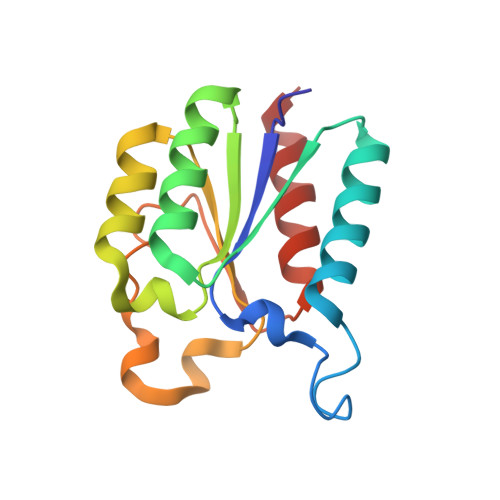 | |
UniProt | |||||
Entity Groups | |||||
| Sequence Clusters | 30% Identity50% Identity70% Identity90% Identity95% Identity100% Identity | ||||
| UniProt Group | A3M692 | ||||
Sequence AnnotationsExpand | |||||
Reference Sequence | |||||
| Length ( Å ) | Angle ( ˚ ) |
|---|---|
| a = 97.914 | α = 90 |
| b = 136.008 | β = 97.59 |
| c = 142.878 | γ = 90 |
| Software Name | Purpose |
|---|---|
| REFMAC | refinement |
| HKL-2000 | data reduction |
| HKL-2000 | data scaling |
| MOLREP | phasing |


