4Z93 | pdb_00004z93

BRD4 bromodomain 2 in complex with gamma-carboline-containing compound, number 18.


Experimental Data Snapshot

  • Method: X-RAY DIFFRACTION
  • Resolution: 1.27 Å
  • R-Value Free: 
    0.184 (Depositor), 0.169 (DCC) 
  • R-Value Work: 
    0.155 (Depositor), 0.149 (DCC) 
  • R-Value Observed: 
    0.157 (Depositor) 

Starting Model: experimental
View more details

wwPDB Validation 3D Report Full Report

Validation slider image for 4Z93

Ligand Structure Quality Assessment 


This is version 1.3 of the entry. See complete history

Literature

Structure-Based Design of gamma-Carboline Analogues as Potent and Specific BET Bromodomain Inhibitors.

Ran, X.Zhao, Y.Liu, L.Bai, L.Yang, C.Y.Zhou, B.Meagher, J.L.Chinnaswamy, K.Stuckey, J.A.Wang, S.

(2015) J Med Chem 58: 4927-4939

  • DOI: https://doi.org/10.1021/acs.jmedchem.5b00613
  • Primary Citation Related Structures: 
    4Z93

  • PubMed Abstract: 

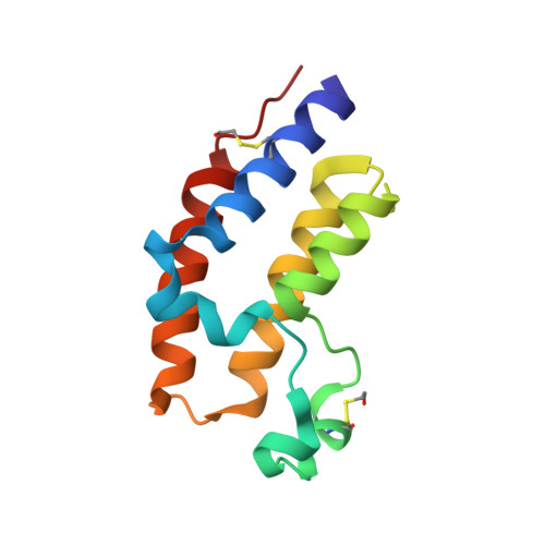
    Small-molecule inhibitors of bromodomain and extra terminal proteins (BET), including BRD2, BRD3, and BRD4 proteins have therapeutic potential for the treatment of human cancers and other diseases and conditions. In this paper, we report the design, synthesis, and evaluation of γ-carboline-containing compounds as a new class of small-molecule BET inhibitors. The most potent inhibitor (compound 18, RX-37) obtained from this study binds to BET bromodomain proteins (BRD2, BRD3, and BRD4) with Ki values of 3.2-24.7 nM and demonstrates high selectivity over other non-BET bromodomain-containing proteins. Compound 18 potently and selectively inhibits cell growth in human acute leukemia cell lines harboring the rearranged mixed lineage leukemia 1 gene. We have determined a cocrystal structure of 18 in complex with BRD4 BD2 at 1.4 Å resolution, which provides a solid structural basis for the compound's high binding affinity and for its further structure-based optimization. Compound 18 represents a promising lead compound for the development of a new class of therapeutics for the treatment of human cancer and other conditions.


  • Organizational Affiliation
    • †Departments of Medicinal Chemistry, ‡Internal Medicine, §Pharmacology, and ∥Biological Chemistry, ⊥Life Sciences Institute, and #Comprehensive Cancer Center, University of Michigan, Ann Arbor, Michigan 48109, United States.

Macromolecule Content 

  • Total Structure Weight: 13.73 kDa 
  • Atom Count: 1,081 
  • Modeled Residue Count: 112 
  • Deposited Residue Count: 112 
  • Unique protein chains: 1

Macromolecules

Find similar proteins by:|  3D Structure
Entity ID: 1
MoleculeChains  Sequence LengthOrganismDetailsImage
Bromodomain-containing protein 4112Homo sapiensMutation(s): 0 
Gene Names: BRD4HUNK1
UniProt & NIH Common Fund Data Resources
Find proteins for O60885 (Homo sapiens)
Explore O60885 
Go to UniProtKB:  O60885
PHAROS:  O60885
GTEx:  ENSG00000141867 
Entity Groups
Sequence Clusters30% Identity50% Identity70% Identity90% Identity95% Identity100% Identity
UniProt GroupO60885
Sequence Annotations
Expand
Reference Sequence

Small Molecules

Ligands 2 Unique
IDChains Name / Formula / InChI Key2D Diagram3D Interactions
4LD

Query on 4LD


Download:Ideal Coordinates CCD File
B [auth A]1-(3-cyclopropyl-5-methyl-1H-pyrazol-4-yl)-7-(3,5-dimethyl-1,2-oxazol-4-yl)-8-methoxy-5H-pyrido[4,3-b]indole
C24 H23 N5 O2
WHDUHJYPRJXLGF-UHFFFAOYSA-N
EDO

Query on EDO


Download:Ideal Coordinates CCD File
C [auth A]1,2-ETHANEDIOL
C2 H6 O2
LYCAIKOWRPUZTN-UHFFFAOYSA-N
Modified Residues  1 Unique
IDChains TypeFormula2D DiagramParent
CME
Query on CME
A
L-PEPTIDE LINKINGC5 H11 N O3 S2CYS

Experimental Data & Validation

Experimental Data

  • Method: X-RAY DIFFRACTION
  • Resolution: 1.27 Å
  • R-Value Free:  0.184 (Depositor), 0.169 (DCC) 
  • R-Value Work:  0.155 (Depositor), 0.149 (DCC) 
  • R-Value Observed: 0.157 (Depositor) 
Space Group: P 21 21 2
Unit Cell:
Length ( Å )Angle ( ˚ )
a = 51.862α = 90
b = 72.696β = 90
c = 32.115γ = 90
Software Package:
Software NamePurpose
HKL-2000data scaling
BUSTER-TNTrefinement
PDB_EXTRACTdata extraction
HKL-2000data reduction
PHASERphasing

Structure Validation

View Full Validation Report


Ligand Structure Quality Assessment 


Entry History 

Deposition Data

Revision History  (Full details and data files)

  • Version 1.0: 2015-07-01
    Type: Initial release
  • Version 1.1: 2015-07-08
    Changes: Experimental preparation, Structure summary
  • Version 1.2: 2023-09-27
    Changes: Advisory, Data collection, Database references, Derived calculations, Refinement description, Source and taxonomy, Structure summary
  • Version 1.3: 2024-10-30
    Changes: Structure summary