4Z8B | pdb_00004z8b

crystal structure of a DGL mutant - H51G H131N


Experimental Data Snapshot

  • Method: X-RAY DIFFRACTION
  • Resolution: 1.95 Å
  • R-Value Free: 
    0.243 (Depositor), 0.251 (DCC) 
  • R-Value Work: 
    0.201 (Depositor), 0.206 (DCC) 
  • R-Value Observed: 
    0.203 (Depositor) 

Starting Model: experimental
View more details

wwPDB Validation 3D Report Full Report

Validation slider image for 4Z8B

Ligand Structure Quality Assessment 


This is version 1.4 of the entry. See complete history

Literature

Quaternary structure of Dioclea grandiflora lectin assessed by equilibrium sedimentation and crystallographic analysis of recombinant mutants.

Zamora-Caballero, S.Perez, A.Sanz, L.Bravo, J.Calvete, J.J.

(2015) FEBS Lett 589: 2290-2296

  • DOI: https://doi.org/10.1016/j.febslet.2015.07.020
  • Primary Citation Related Structures: 
    4Z8B

  • PubMed Abstract: 

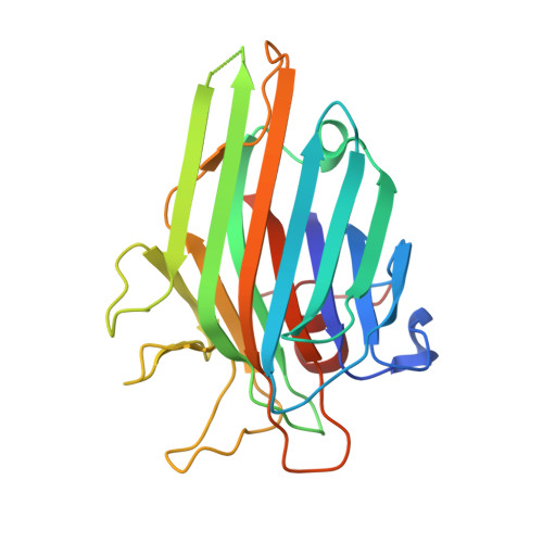
    The structural basis of the pH dependency of the dimer-tetramer transition exhibited by Brinda's type II Diocleinae lectins was investigated by equilibrium sedimentation and X-ray crystal structure determination of recombinant wild-type and site-directed single and double mutants of the pH-stable tetrameric Dioclea grandiflora lectin (r-αDGL). Releasing the peripheral site interdimeric contact between R60 and D78 rendered a mutant displaying dimer-tetramer equilibrium in the pH range equivalent to pKa±1 of the γ-COOH. Mutation of both histidines 51 and 131, but not the mutation of each His separately, abolished the formation of the Diocleinae canonical tetramer in the pH range 2.5-8.5. The X-ray structure of the double mutant r-αDGL H51G/H131N suggests that H131 plays a crucial role in networking loop 114-125 residues from all four subunits at the central cavity of the tetrameric lectin, and that H51 maintains the central cavity loops in a proper spatial orientation to make H131-mediated interdimer contacts.


  • Organizational Affiliation
    • Unidad de Transducción de Señales, Instituto de Biomedicina de Valencia, CSIC, Spain.

Macromolecule Content 

  • Total Structure Weight: 26.31 kDa 
  • Atom Count: 1,893 
  • Modeled Residue Count: 231 
  • Deposited Residue Count: 237 
  • Unique protein chains: 1

Macromolecules

Find similar proteins by:|  3D Structure
Entity ID: 1
MoleculeChains  Sequence LengthOrganismDetailsImage
Lectin alpha chain237Macropsychanthus grandiflorusMutation(s): 0 
UniProt
Find proteins for P08902 (Dioclea grandiflora)
Explore P08902 
Go to UniProtKB:  P08902
Entity Groups
Sequence Clusters30% Identity50% Identity70% Identity90% Identity95% Identity100% Identity
UniProt GroupP08902
Sequence Annotations
Expand
Reference Sequence

Small Molecules

Ligands 5 Unique
IDChains Name / Formula / InChI Key2D Diagram3D Interactions
XMM

Query on XMM


Download:Ideal Coordinates CCD File
G [auth A]5-bromo-4-chloro-1H-indol-3-yl alpha-D-mannopyranoside
C14 H15 Br Cl N O6
OPIFSICVWOWJMJ-HAAGFXOZSA-N
SO4

Query on SO4


Download:Ideal Coordinates CCD File
D [auth A],
E [auth A]
SULFATE ION
O4 S
QAOWNCQODCNURD-UHFFFAOYSA-L
GOL

Query on GOL


Download:Ideal Coordinates CCD File
F [auth A]GLYCEROL
C3 H8 O3
PEDCQBHIVMGVHV-UHFFFAOYSA-N
MN

Query on MN


Download:Ideal Coordinates CCD File
C [auth A]MANGANESE (II) ION
Mn
WAEMQWOKJMHJLA-UHFFFAOYSA-N
CA

Query on CA


Download:Ideal Coordinates CCD File
B [auth A]CALCIUM ION
Ca
BHPQYMZQTOCNFJ-UHFFFAOYSA-N

Experimental Data & Validation

Experimental Data

  • Method: X-RAY DIFFRACTION
  • Resolution: 1.95 Å
  • R-Value Free:  0.243 (Depositor), 0.251 (DCC) 
  • R-Value Work:  0.201 (Depositor), 0.206 (DCC) 
  • R-Value Observed: 0.203 (Depositor) 
Space Group: I 2 2 2
Unit Cell:
Length ( Å )Angle ( ˚ )
a = 62.35α = 90
b = 67.08β = 90
c = 108.62γ = 90
Software Package:
Software NamePurpose
PHENIXrefinement
iMOSFLMdata reduction
SCALAdata scaling
PHENIXphasing

Structure Validation

View Full Validation Report


Ligand Structure Quality Assessment 


Entry History 

Deposition Data

Revision History  (Full details and data files)

  • Version 1.0: 2015-07-22
    Type: Initial release
  • Version 1.1: 2015-08-12
    Changes: Database references
  • Version 1.2: 2015-08-19
    Changes: Database references
  • Version 1.3: 2020-07-29
    Type: Remediation
    Reason: Carbohydrate remediation
    Changes: Data collection, Derived calculations, Structure summary
  • Version 1.4: 2024-01-10
    Changes: Data collection, Database references, Refinement description, Structure summary