4Z76 | pdb_00004z76

Weak TCR binding to an unstable insulin epitope drives type 1 diabetes


Experimental Data Snapshot

  • Method: X-RAY DIFFRACTION
  • Resolution: 1.88 Å
  • R-Value Free: 
    0.230 (Depositor), 0.235 (DCC) 
  • R-Value Work: 
    0.184 (Depositor), 0.191 (DCC) 
  • R-Value Observed: 
    0.186 (Depositor) 

wwPDB Validation 3D Report Full Report

Validation slider image for 4Z76

This is version 1.3 of the entry. See complete history

Literature

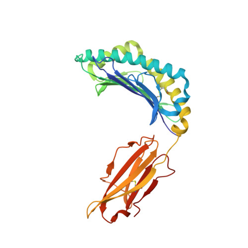
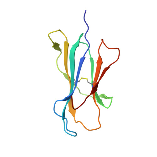
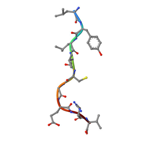
Distortion of the Major Histocompatibility Complex Class I Binding Groove to Accommodate an Insulin-derived 10-Mer Peptide.

Motozono, C.Pearson, J.A.De Leenheer, E.Rizkallah, P.J.Beck, K.Trimby, A.Sewell, A.K.Wong, F.S.Cole, D.K.

(2015) J Biological Chem 290: 18924-18933

  • DOI: https://doi.org/10.1074/jbc.M114.622522
  • Primary Citation Related Structures: 
    4WDI, 4Z76, 4Z78

  • PubMed Abstract: 

    The non-obese diabetic mouse model of type 1 diabetes continues to be an important tool for delineating the role of T-cell-mediated destruction of pancreatic β-cells. However, little is known about the molecular mechanisms that enable this disease pathway. We show that insulin reactivity by a CD8(+) T-cell clone, known to induce type 1 diabetes, is characterized by weak T-cell antigen receptor binding to a relatively unstable peptide-MHC. The structure of the native 9- and 10-mer insulin epitopes demonstrated that peptide residues 7 and 8 form a prominent solvent-exposed bulge that could potentially be the main focus of T-cell receptor binding. The C terminus of the peptide governed peptide-MHC stability. Unexpectedly, we further demonstrate a novel mode of flexible peptide presentation in which the MHC peptide-binding groove is able to "open the back door" to accommodate extra C-terminal peptide residues.


  • Organizational Affiliation
    • From the Division of Infection and Immunity and the Department of Immunology, Kinki University School of Medicine, Osaka 589-8511, Japan, and.

Macromolecule Content 

  • Total Structure Weight: 92.66 kDa 
  • Atom Count: 7,048 
  • Modeled Residue Count: 772 
  • Deposited Residue Count: 772 
  • Unique protein chains: 3

Macromolecules

Find similar proteins by:|  3D Structure
Entity ID: 1
MoleculeChains  Sequence LengthOrganismDetailsImage
H-2 class I histocompatibility antigen, K-D alpha chain
A, D
277Mus musculusMutation(s): 0 
Gene Names: H2-K1H2-K
UniProt
Find proteins for P01902 (Mus musculus)
Explore P01902 
Go to UniProtKB:  P01902
Entity Groups
Sequence Clusters30% Identity50% Identity70% Identity90% Identity95% Identity100% Identity
UniProt GroupP01902
Sequence Annotations
Expand
Reference Sequence
Find similar proteins by:|  3D Structure
Entity ID: 2
MoleculeChains  Sequence LengthOrganismDetailsImage
Beta-2-microglobulin
B, E
100Homo sapiensMutation(s): 0 
Gene Names: B2MCDABP0092HDCMA22P
UniProt & NIH Common Fund Data Resources
Find proteins for P61769 (Homo sapiens)
Explore P61769 
Go to UniProtKB:  P61769
PHAROS:  P61769
GTEx:  ENSG00000166710 
Entity Groups
Sequence Clusters30% Identity50% Identity70% Identity90% Identity95% Identity100% Identity
UniProt GroupP61769
Sequence Annotations
Expand
Reference Sequence
Find similar proteins by:  Sequence   |   3D Structure  
Entity ID: 3
MoleculeChains  Sequence LengthOrganismDetailsImage
Insulin
C, F
9Homo sapiensMutation(s): 0 
Gene Names: INS
UniProt & NIH Common Fund Data Resources
Find proteins for P01308 (Homo sapiens)
Explore P01308 
Go to UniProtKB:  P01308
PHAROS:  P01308
GTEx:  ENSG00000254647 
Entity Groups
UniProt GroupP01308
Sequence Annotations
Expand
Reference Sequence

Small Molecules

Ligands 3 Unique
IDChains Name / Formula / InChI Key2D Diagram3D Interactions
SO4

Query on SO4


Download:Ideal Coordinates CCD File
AA [auth E]
BA [auth E]
CA [auth E]
DA [auth E]
K [auth A]
AA [auth E],
BA [auth E],
CA [auth E],
DA [auth E],
K [auth A],
O [auth B],
P [auth B],
Q [auth B],
R [auth D],
S [auth D],
T [auth D],
U [auth D],
V [auth D],
W [auth D],
Z [auth E]
SULFATE ION
O4 S
QAOWNCQODCNURD-UHFFFAOYSA-L
GOL

Query on GOL


Download:Ideal Coordinates CCD File
I [auth A],
J [auth A],
Y [auth E]
GLYCEROL
C3 H8 O3
PEDCQBHIVMGVHV-UHFFFAOYSA-N
EDO

Query on EDO


Download:Ideal Coordinates CCD File
G [auth A]
H [auth A]
L [auth B]
M [auth B]
N [auth B]
G [auth A],
H [auth A],
L [auth B],
M [auth B],
N [auth B],
X [auth E]
1,2-ETHANEDIOL
C2 H6 O2
LYCAIKOWRPUZTN-UHFFFAOYSA-N

Experimental Data & Validation

Experimental Data

  • Method: X-RAY DIFFRACTION
  • Resolution: 1.88 Å
  • R-Value Free:  0.230 (Depositor), 0.235 (DCC) 
  • R-Value Work:  0.184 (Depositor), 0.191 (DCC) 
  • R-Value Observed: 0.186 (Depositor) 
Space Group: P 1
Unit Cell:
Length ( Å )Angle ( ˚ )
a = 47.99α = 69.81
b = 62.37β = 85.76
c = 72.31γ = 87.05
Software Package:
Software NamePurpose
REFMACrefinement
xia2data reduction
SCALAdata scaling
PHASERphasing

Structure Validation

View Full Validation Report


Entry History 

Deposition Data

Revision History  (Full details and data files)

  • Version 1.0: 2015-06-24
    Type: Initial release
  • Version 1.1: 2015-07-01
    Changes: Database references
  • Version 1.2: 2015-08-12
    Changes: Database references
  • Version 1.3: 2024-10-23
    Changes: Data collection, Database references, Refinement description, Structure summary