4Z2P

Crystal structure of short hoefavidin-hoef-peptide(L9F) complex


Experimental Data Snapshot

  • Method: X-RAY DIFFRACTION
  • Resolution: 1.60 Å
  • R-Value Free: 0.207 
  • R-Value Work: 0.172 
  • R-Value Observed: 0.173 

wwPDB Validation   3D Report Full Report


This is version 1.4 of the entry. See complete history


Literature

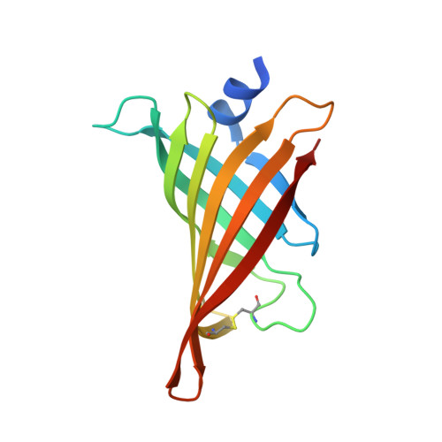
Hoefavidin: A dimeric bacterial avidin with a C-terminal binding tail.

Avraham, O.Meir, A.Fish, A.Bayer, E.A.Livnah, O.

(2015) J Struct Biol 191: 139-148

  • DOI: https://doi.org/10.1016/j.jsb.2015.06.020
  • Primary Citation of Related Structures:  
    4Z27, 4Z28, 4Z2O, 4Z2P, 4Z2V, 4Z6J

  • PubMed Abstract: 

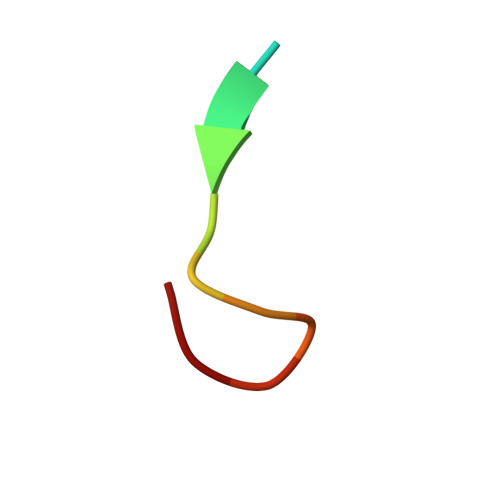
    Dimeric avidins are a newly discovered subgroup of the avidin family that bind biotin with high affinity. Their dimeric configuration is a quaternary substructure of the classical tetrameric avidins which lacks the requirement of the critical Trp that defines the tetramer and dictates the tenacious interaction with biotin. Hoefavidin, derived from the bacterium Hoeflea phototrophica DFL-43(T), is the third characterized member of the dimeric avidin subfamily. Like the other members of this group, hoefavidin is a thermostable protein that contains a disulfide bridge between Cys57 and Cys88, thereby connecting and stabilizing the L3,4 and L5,6 loops. This represents a distinctive characteristic of dimeric avidins that compensates for the lack of Trp and enables their dimeric configuration. The X-ray structure of the intact hoefavidin revealed unique crystal packing generated by an octameric cylindrical structure wherein the C-termini segments of each monomer is introduced into the entrance of the biotin-binding site of an adjacent non-canonical monomer. This anomaly in the protein structure served as a lead toward the design of specific binding peptides. We screened for specific hoefavidin binding peptides derived from the C-terminal region and two peptides were obtained that bind a truncated form of hoefavidin (lacking the last 10 amino acids) with dissociation constants of 10(-5)M. The crystal structure of short hoefavidin complexed with a C-terminal derived peptide revealed the mode of binding. These peptides may form the basis of novel and reversible binders for dimeric avidins.


  • Organizational Affiliation

    Department of Biological Chemistry, The Alexander Silberman Institute of Life Sciences, The Wolfson Centre for Applied Structural Biology, The Hebrew University of Jerusalem, The Edmond J. Safra Campus, Jerusalem 91904, Israel.


Macromolecules
Find similar proteins by:  (by identity cutoff)  |  3D Structure
Entity ID: 1
MoleculeChains Sequence LengthOrganismDetailsImage
Avidin familyA,
C [auth B]
134Hoeflea phototrophica DFL-43Mutation(s): 0 
Gene Names: HPDFL43_17171
UniProt
Find proteins for A9D857 (Hoeflea phototrophica (strain DSM 17068 / NCIMB 14078 / DFL-43))
Explore A9D857 
Go to UniProtKB:  A9D857
Entity Groups  
Sequence Clusters30% Identity50% Identity70% Identity90% Identity95% Identity100% Identity
UniProt GroupA9D857
Sequence Annotations
Expand
  • Reference Sequence

Find similar proteins by:  Sequence   |   3D Structure  

Entity ID: 2
MoleculeChains Sequence LengthOrganismDetailsImage
Hoef-peptide (L9F)B [auth P],
D [auth C]
12Hoeflea phototrophica DFL-43Mutation(s): 0 
UniProt
Find proteins for A9D857 (Hoeflea phototrophica (strain DSM 17068 / NCIMB 14078 / DFL-43))
Explore A9D857 
Go to UniProtKB:  A9D857
Entity Groups  
Sequence Clusters30% Identity50% Identity70% Identity90% Identity95% Identity100% Identity
UniProt GroupA9D857
Sequence Annotations
Expand
  • Reference Sequence
Experimental Data & Validation

Experimental Data

  • Method: X-RAY DIFFRACTION
  • Resolution: 1.60 Å
  • R-Value Free: 0.207 
  • R-Value Work: 0.172 
  • R-Value Observed: 0.173 
  • Space Group: P 43 21 2
Unit Cell:
Length ( Å )Angle ( ˚ )
a = 61.228α = 90
b = 61.228β = 90
c = 166.817γ = 90
Software Package:
Software NamePurpose
REFMACrefinement
MOSFLMdata reduction
MOLREPphasing
SCALEPACKdata scaling

Structure Validation

View Full Validation Report


Entry History 

Deposition Data

Revision History  (Full details and data files)

  • Version 1.0: 2015-07-15
    Type: Initial release
  • Version 1.1: 2015-08-05
    Changes: Database references
  • Version 1.2: 2017-11-22
    Changes: Database references
  • Version 1.3: 2019-09-18
    Changes: Data collection
  • Version 1.4: 2024-01-10
    Changes: Data collection, Database references, Refinement description