4YXP | pdb_00004yxp

The structure of the folded domain of the signature multifunctional protein ICP27 from herpes simplex virus-1 reveals an intertwined dimer.


Experimental Data Snapshot

  • Method: X-RAY DIFFRACTION
  • Resolution: 1.92 Å
  • R-Value Free: 
    0.203 (Depositor), 0.213 (DCC) 
  • R-Value Work: 
    0.168 (Depositor), 0.180 (DCC) 
  • R-Value Observed: 
    0.170 (Depositor) 

wwPDB Validation 3D Report Full Report

Validation slider image for 4YXP

This is version 1.2 of the entry. See complete history

Literature

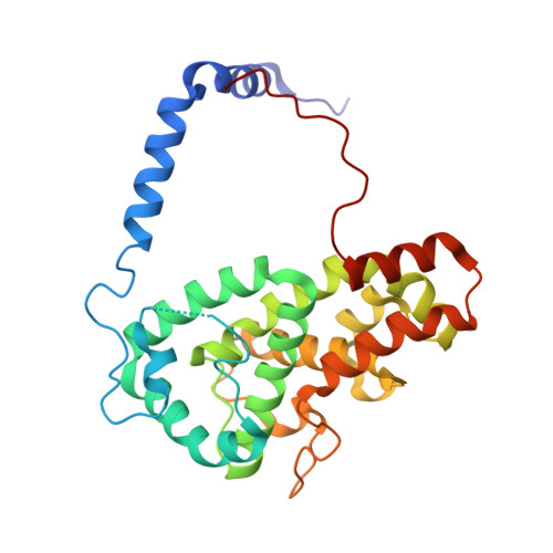
The structure of the folded domain from the signature multifunctional protein ICP27 from herpes simplex virus-1 reveals an intertwined dimer.

Tunnicliffe, R.B.Schacht, M.Levy, C.Jowitt, T.A.Sandri-Goldin, R.M.Golovanov, A.P.

(2015) Sci Rep 5: 11234-11234

  • DOI: https://doi.org/10.1038/srep11234
  • Primary Citation Related Structures: 
    4YXP

  • PubMed Abstract: 

    Herpesviruses cause life-long infections by evading the host immune system and establishing latent infections. All mammalian herpesviruses express an essential multifunctional protein that is typified by ICP27 encoded by Herpes Simplex Virus 1. The only region that is conserved among the diverse members of the ICP27 family is a predicted globular domain that has been termed the ICP27 homology domain. Here we present the first crystal structure of the ICP27 homology domain, solved to 1.9 Å resolution. The protein is a homo-dimer, adopting a novel intertwined fold with one CHCC zinc-binding site per monomer. The dimerization, which was independently confirmed by SEC-MALS and AUC, is stabilized by an extensive network of intermolecular contacts, and a domain-swap involving the two N-terminal helices and C-terminal tails. Each monomer contains a lid motif that can clamp the C-terminal tail of its dimeric binding partner against its globular core, without forming any distinct secondary structure elements. The binding interface was probed with point mutations, none of which had a noticeable effect on dimer formation; however deletion of the C-terminal tail region prevented dimer formation in vivo. The structure provides a template for future biochemical studies and modelling of ICP27 homologs from other herpesviruses.


  • Organizational Affiliation
    • Manchester Institute of Biotechnology, The University of Manchester, Manchester, UK.

Macromolecule Content 

  • Total Structure Weight: 60.33 kDa 
  • Atom Count: 4,512 
  • Modeled Residue Count: 539 
  • Deposited Residue Count: 550 
  • Unique protein chains: 1

Macromolecules

Find similar proteins by:|  3D Structure
Entity ID: 1
MoleculeChains  Sequence LengthOrganismDetailsImage
mRNA export factor
A, B
275Human alphaherpesvirus 1 strain 17Mutation(s): 0 
Gene Names: UL54
UniProt
Find proteins for P10238 (Human herpesvirus 1 (strain 17))
Explore P10238 
Go to UniProtKB:  P10238
Entity Groups
Sequence Clusters30% Identity50% Identity70% Identity90% Identity95% Identity100% Identity
UniProt GroupP10238
Sequence Annotations
Expand
Reference Sequence

Experimental Data & Validation

Experimental Data

  • Method: X-RAY DIFFRACTION
  • Resolution: 1.92 Å
  • R-Value Free:  0.203 (Depositor), 0.213 (DCC) 
  • R-Value Work:  0.168 (Depositor), 0.180 (DCC) 
  • R-Value Observed: 0.170 (Depositor) 
Space Group: P 21 21 2
Unit Cell:
Length ( Å )Angle ( ˚ )
a = 99.33α = 90
b = 133.62β = 90
c = 44.97γ = 90
Software Package:
Software NamePurpose
PHENIXrefinement
xia2data reduction
xia2data scaling
PHENIXphasing

Structure Validation

View Full Validation Report


Entry History 

& Funding Information

Deposition Data


Funding OrganizationLocationGrant Number
National Institutes of HealthUnited StatesAI107803

Revision History  (Full details and data files)

  • Version 1.0: 2015-06-17
    Type: Initial release
  • Version 1.1: 2015-06-24
    Changes: Database references
  • Version 1.2: 2024-05-08
    Changes: Data collection, Database references