4YT1 | pdb_00004yt1

Human PPAR Gamma Ligand Binding Domain in complex with a Gammma Selective Synthetic Partial Agonist MEKT76


Experimental Data Snapshot

  • Method: X-RAY DIFFRACTION
  • Resolution: 2.20 Å
  • R-Value Free: 
    0.256 (Depositor), 0.258 (DCC) 
  • R-Value Work: 
    0.237 (Depositor), 0.239 (DCC) 
  • R-Value Observed: 
    0.238 (Depositor) 

Starting Model: experimental
View more details

wwPDB Validation 3D Report Full Report

Validation slider image for 4YT1

Ligand Structure Quality Assessment 


This is version 1.2 of the entry. See complete history

Literature

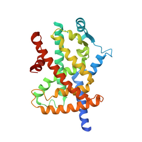
Peroxisome proliferator-activated receptor gamma (PPAR gamma ) has multiple binding points that accommodate ligands in various conformations: Structurally similar PPAR gamma partial agonists bind to PPAR gamma LBD in different conformations

Ohashi, M.Gamo, K.Oyama, T.Miyachi, H.

(2015) Bioorg Med Chem Lett 25: 2758-2762

  • DOI: https://doi.org/10.1016/j.bmcl.2015.05.025
  • Primary Citation Related Structures: 
    4YT1

  • PubMed Abstract: 

    In the course of studies directed toward the creation of human peroxisome proliferator-activated receptor gamma (hPPARγ) partial agonists, we designed and synthesized benzylsulfonylaminocarbonyl derivative (3) by structural modification of our reported hPPARγ partial agonist 2. Co-crystallization of 3 with the hPPARγ ligand-binding domain (LBD) afforded a homodimeric complex in which one of the LBDs adopts a fully active structure without bound 3, while the other LBD exhibits a non-fully active structure containing one molecule of bound 3. Interestingly, 2 and 3 are structurally similar, but bind to hPPARγ LBD in distinct conformations, that is, the sulfonylaminocarbonyl moiety of bound 3 is directed at 180° away from that of bound 2. These results support our previous proposal that the hPPARγ LBD has multiple binding points that can be utilized to accommodate structurally flexible hPPAR ligands.


  • Organizational Affiliation
    • Graduate School of Medicine, Dentistry and Pharmaceutical Sciences, Okayama University, 1-1-1, Tsushima-Naka, Kita-ku, Okayama 700-8530, Japan.

Macromolecule Content 

  • Total Structure Weight: 65.61 kDa 
  • Atom Count: 4,199 
  • Modeled Residue Count: 518 
  • Deposited Residue Count: 572 
  • Unique protein chains: 1

Macromolecules

Find similar proteins by:|  3D Structure
Entity ID: 1
MoleculeChains  Sequence LengthOrganismDetailsImage
Peroxisome proliferator-activated receptor gamma
A, B
286Homo sapiensMutation(s): 0 
Gene Names: PPARGNR1C3
UniProt & NIH Common Fund Data Resources
Find proteins for P37231 (Homo sapiens)
Explore P37231 
Go to UniProtKB:  P37231
PHAROS:  P37231
GTEx:  ENSG00000132170 
Entity Groups
Sequence Clusters30% Identity50% Identity70% Identity90% Identity95% Identity100% Identity
UniProt GroupP37231
Sequence Annotations
Expand
Reference Sequence

Small Molecules

Ligands 1 Unique
IDChains Name / Formula / InChI Key2D Diagram3D Interactions
JJB

Query on JJB


Download:Ideal Coordinates CCD File
C [auth B]N-(benzylsulfonyl)-4-propoxy-3-({[4-(pyrimidin-2-yl)benzoyl]amino}methyl)benzamide
C29 H28 N4 O5 S
JNDTXHSMALXHGN-UHFFFAOYSA-N

Experimental Data & Validation

Experimental Data

  • Method: X-RAY DIFFRACTION
  • Resolution: 2.20 Å
  • R-Value Free:  0.256 (Depositor), 0.258 (DCC) 
  • R-Value Work:  0.237 (Depositor), 0.239 (DCC) 
  • R-Value Observed: 0.238 (Depositor) 
Space Group: C 1 2 1
Unit Cell:
Length ( Å )Angle ( ˚ )
a = 93.279α = 90
b = 60.815β = 103.74
c = 118.936γ = 90
Software Package:
Software NamePurpose
PHENIXrefinement
HKL-2000data reduction
SCALEPACKdata scaling
PHENIXphasing

Structure Validation

View Full Validation Report


Ligand Structure Quality Assessment 


Entry History 

Deposition Data

Revision History  (Full details and data files)

  • Version 1.0: 2016-03-02
    Type: Initial release
  • Version 1.1: 2020-02-05
    Changes: Data collection, Derived calculations
  • Version 1.2: 2023-11-08
    Changes: Data collection, Database references, Refinement description