4YMA | pdb_00004yma

Structure of the ligand-binding domain of GluA2 in complex with the antagonist CNG10109


Experimental Data Snapshot

  • Method: X-RAY DIFFRACTION
  • Resolution: 1.90 Å
  • R-Value Free: 
    0.228 (Depositor), 0.230 (DCC) 
  • R-Value Work: 
    0.170 (Depositor), 0.174 (DCC) 
  • R-Value Observed: 
    0.173 (Depositor) 

Starting Model: experimental
View more details

wwPDB Validation 3D Report Full Report

Validation slider image for 4YMA

Ligand Structure Quality Assessment 


This is version 1.5 of the entry. See complete history

Literature

Structure-Activity Relationship Study of Ionotropic Glutamate Receptor Antagonist (2S,3R)-3-(3-Carboxyphenyl)pyrrolidine-2-carboxylic Acid.

Krogsgaard-Larsen, N.Storgaard, M.Moller, C.Demmer, C.S.Hansen, J.Han, L.Monrad, R.N.Nielsen, B.Tapken, D.Pickering, D.S.Kastrup, J.S.Frydenvang, K.Bunch, L.

(2015) J Med Chem 58: 6131-6150

  • DOI: https://doi.org/10.1021/acs.jmedchem.5b00750
  • Primary Citation Related Structures: 
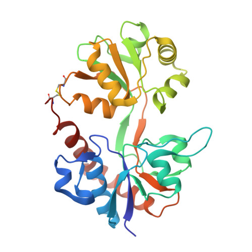
    4YMA, 4YMB

  • PubMed Abstract: 

    Herein we describe the first structure-activity relationship study of the broad-range iGluR antagonist (2S,3R)-3-(3-carboxyphenyl)pyrrolidine-2-carboxylic acid (1) by exploring the pharmacological effect of substituents in the 4, 4', or 5' positions and the bioisosteric substitution of the distal carboxylic acid for a phosphonic acid moiety. Of particular interest is a hydroxyl group in the 4' position 2a which induced a preference in binding affinity for homomeric GluK3 over GluK1 (Ki = 0.87 and 4.8 μM, respectively). Two X-ray structures of ligand binding domains were obtained: 2e in GluA2-LBD and 2f in GluK1-LBD, both at 1.9 Å resolution. Compound 2e induces a D1-D2 domain opening in GluA2-LBD of 17.3-18.8° and 2f a domain opening in GluK1-LBD of 17.0-17.5° relative to the structures with glutamate. The pyrrolidine-2-carboxylate moiety of 2e and 2f shows a similar binding mode as kainate. The 3-carboxyphenyl ring of 2e and 2f forms contacts comparable to those of the distal carboxylate in kainate.


  • Organizational Affiliation
    • †Chemical Neuroscience Group, ‡Biostructural Research Group, §Medicinal Chemistry Group, ∥Molecular, Cellular Pharmacology Group, Department of Drug Design and Pharmacology, Faculty of Health and Medical Sciences, University of Copenhagen, Universitetsparken 2, 2100 Copenhagen Ø, Denmark.

Macromolecule Content 

  • Total Structure Weight: 60 kDa 
  • Atom Count: 4,898 
  • Modeled Residue Count: 518 
  • Deposited Residue Count: 528 
  • Unique protein chains: 1

Macromolecules

Find similar proteins by:|  3D Structure
Entity ID: 1
MoleculeChains  Sequence LengthOrganismDetailsImage
Glutamate receptor 2A [auth B],
B [auth A]
264Rattus norvegicusMutation(s): 0 
Gene Names: Gria2Glur2
UniProt
Find proteins for P19491 (Rattus norvegicus)
Explore P19491 
Go to UniProtKB:  P19491
Entity Groups
Sequence Clusters30% Identity50% Identity70% Identity90% Identity95% Identity100% Identity
UniProt GroupP19491
Sequence Annotations
Expand
Reference Sequence

Small Molecules

Ligands 5 Unique
IDChains Name / Formula / InChI Key2D Diagram3D Interactions
4E5

Query on 4E5


Download:Ideal Coordinates CCD File
F [auth B],
O [auth A]
(3R)-3-(3-carboxy-5-hydroxyphenyl)-L-proline
C12 H13 N O5
RMJSXKKIOKMPOU-ZJUUUORDSA-N
SO4

Query on SO4


Download:Ideal Coordinates CCD File
C [auth B]
D [auth B]
J [auth A]
K [auth A]
L [auth A]
C [auth B],
D [auth B],
J [auth A],
K [auth A],
L [auth A],
M [auth A]
SULFATE ION
O4 S
QAOWNCQODCNURD-UHFFFAOYSA-L
GOL

Query on GOL


Download:Ideal Coordinates CCD File
E [auth B],
N [auth A]
GLYCEROL
C3 H8 O3
PEDCQBHIVMGVHV-UHFFFAOYSA-N
EDO

Query on EDO


Download:Ideal Coordinates CCD File
G [auth B],
H [auth B]
1,2-ETHANEDIOL
C2 H6 O2
LYCAIKOWRPUZTN-UHFFFAOYSA-N
ACT

Query on ACT


Download:Ideal Coordinates CCD File
I [auth A]ACETATE ION
C2 H3 O2
QTBSBXVTEAMEQO-UHFFFAOYSA-M

Experimental Data & Validation

Experimental Data

  • Method: X-RAY DIFFRACTION
  • Resolution: 1.90 Å
  • R-Value Free:  0.228 (Depositor), 0.230 (DCC) 
  • R-Value Work:  0.170 (Depositor), 0.174 (DCC) 
  • R-Value Observed: 0.173 (Depositor) 
Space Group: P 1 21 1
Unit Cell:
Length ( Å )Angle ( ˚ )
a = 48.88α = 90
b = 65.25β = 92.36
c = 91.55γ = 90
Software Package:
Software NamePurpose
PHENIXrefinement
SCALAdata scaling
PHASERphasing
PDB_EXTRACTdata extraction
MOSFLMdata reduction
Cootmodel building

Structure Validation

View Full Validation Report


Ligand Structure Quality Assessment 


Entry History 

& Funding Information

Deposition Data


Funding OrganizationLocationGrant Number
Denmark--

Revision History  (Full details and data files)

  • Version 1.0: 2015-08-05
    Type: Initial release
  • Version 1.1: 2015-08-26
    Changes: Database references
  • Version 1.2: 2017-08-09
    Changes: Data collection, Database references
  • Version 1.3: 2018-03-07
    Changes: Source and taxonomy
  • Version 1.4: 2024-01-10
    Changes: Data collection, Database references, Refinement description
  • Version 1.5: 2024-10-23
    Changes: Structure summary