4YM2 | pdb_00004ym2

Crystal structure of the human galectin-4 C-terminal carbohydrate recognition domain in complex with lactose-3'-sulfate


Experimental Data Snapshot

  • Method: X-RAY DIFFRACTION
  • Resolution: 2.10 Å
  • R-Value Free: 
    0.210 (Depositor), 0.216 (DCC) 
  • R-Value Work: 
    0.161 (Depositor), 0.170 (DCC) 
  • R-Value Observed: 
    0.163 (Depositor) 

Starting Model: experimental
View more details

wwPDB Validation 3D Report Full Report

Validation slider image for 4YM2

This is version 2.1 of the entry. See complete history

Literature

Structural characterization of human galectin-4 C-terminal domain: elucidating the molecular basis for recognition of glycosphingolipids, sulfated saccharides and blood group antigens.

Bum-Erdene, K.Leffler, H.Nilsson, U.J.Blanchard, H.

(2015) FEBS J 282: 3348-3367

  • DOI: https://doi.org/10.1111/febs.13348
  • Primary Citation Related Structures: 
    4YLZ, 4YM0, 4YM1, 4YM2, 4YM3

  • PubMed Abstract: 

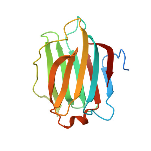
    Human galectin-4 is a lectin that is expressed mainly in the gastrointestinal tract and exhibits metastasis-promoting roles in some cancers. Its tandem-repeat nature exhibits two distinct carbohydrate recognition domains allowing crosslinking by simultaneous binding to sulfated and non-sulfated (but not sialylated) glycosphingolipids and glycoproteins, facilitating stabilization of lipid rafts. Critically, galectin-4 exerts favourable or unfavourable effects depending upon the cancer. Here we report the first X-ray crystallographic structural information on human galectin-4, specifically the C-terminal carbohydrate recognition domain of human (galectin-4C) in complex with lactose, lactose-3'-sulfate, 2'-fucosyllactose, lacto-N-tetraose and lacto-N-neotetraose. These structures enable elucidation of galectin-4C binding fine-specificity towards sulfated and non-sulfated lacto- and neolacto-series sphingolipids as well as to human blood group antigens. Analysis of the lactose-3'-sulfate complex structure shows that galectin-4C does not recognize the sulfate group using any specific amino acid, but binds the ligand nonetheless. Complex structures with lacto-N-tetraose and lacto-N-neotetraose displayed differences in binding interactions exhibited by the non-reducing-end galactose. That of lacto-N-tetraose points outward from the protein surface whereas that of lacto-N-neotetraose interacts directly with the protein. Recognition patterns of human galectin-4C towards lacto- and neolacto-series glycosphingolipids are similar to those of human galectin-3; however, detailed scrutiny revealed differences stemming from the extended binding site that offer distinction in ligand profiles of these two galectins. Structural characterization of the complex with 2'-fucosyllactose, a carbohydrate with similarity to the H antigen, and molecular dynamics studies highlight structural features that allow specific recognition of A and B antigens, whilst a lack of interaction with the 2'-fucose of blood group antigens was revealed. 4YLZ, 4YM0, 4YM1, 4YM2, 4YM3.


  • Organizational Affiliation
    • Institute for Glycomics, Griffith University, Australia.

Macromolecule Content 

  • Total Structure Weight: 69.38 kDa 
  • Atom Count: 4,599 
  • Modeled Residue Count: 556 
  • Deposited Residue Count: 612 
  • Unique protein chains: 1

Macromolecules

Find similar proteins by:|  3D Structure
Entity ID: 1
MoleculeChains  Sequence LengthOrganismDetailsImage
Galectin-4
A, B, C, D
153Homo sapiensMutation(s): 0 
Gene Names: LGALS4
UniProt & NIH Common Fund Data Resources
Find proteins for P56470 (Homo sapiens)
Explore P56470 
Go to UniProtKB:  P56470
PHAROS:  P56470
GTEx:  ENSG00000171747 
Entity Groups
Sequence Clusters30% Identity50% Identity70% Identity90% Identity95% Identity100% Identity
UniProt GroupP56470
Sequence Annotations
Expand
Reference Sequence

Oligosaccharides

Help  
Entity ID: 2
MoleculeChains Length2D Diagram GlycosylationD Interactions
3-O-sulfo-beta-D-galactopyranose-(1-4)-beta-D-glucopyranose
E, F
2N/A
Glycosylation Resources
GlyTouCan: G20044TD
GlyCosmos: G20044TD
GlyGen: G20044TD
Entity ID: 3
MoleculeChains Length2D Diagram GlycosylationD Interactions
beta-D-galactopyranose-(1-4)-beta-D-glucopyranose
G, H
2N/A
Glycosylation Resources
GlyTouCan: G84224TW
GlyCosmos: G84224TW
GlyGen: G84224TW

Experimental Data & Validation

Experimental Data

  • Method: X-RAY DIFFRACTION
  • Resolution: 2.10 Å
  • R-Value Free:  0.210 (Depositor), 0.216 (DCC) 
  • R-Value Work:  0.161 (Depositor), 0.170 (DCC) 
  • R-Value Observed: 0.163 (Depositor) 
Space Group: P 1 21 1
Unit Cell:
Length ( Å )Angle ( ˚ )
a = 45.224α = 90
b = 129.208β = 96.88
c = 45.233γ = 90
Software Package:
Software NamePurpose
REFMACrefinement
Aimlessdata scaling
PDB_EXTRACTdata extraction
XDSdata reduction
PHASERphasing

Structure Validation

View Full Validation Report


Entry History 

& Funding Information

Deposition Data


Funding OrganizationLocationGrant Number
Cancer Council QueenslandAustralia1043716

Revision History  (Full details and data files)

  • Version 1.0: 2015-07-01
    Type: Initial release
  • Version 1.1: 2015-09-09
    Changes: Database references
  • Version 2.0: 2020-07-29
    Type: Remediation
    Reason: Carbohydrate remediation
    Changes: Atomic model, Data collection, Derived calculations, Non-polymer description, Structure summary
  • Version 2.1: 2023-09-27
    Changes: Data collection, Database references, Refinement description, Structure summary