4YKL | pdb_00004ykl

Hnt3 in complex with DNA and guanosine


Experimental Data Snapshot

  • Method: X-RAY DIFFRACTION
  • Resolution: 2.25 Å
  • R-Value Free: 
    0.240 (Depositor), 0.246 (DCC) 
  • R-Value Work: 
    0.180 (Depositor), 0.186 (DCC) 
  • R-Value Observed: 
    0.184 (Depositor) 

Starting Model: experimental
View more details

wwPDB Validation 3D Report Full Report

Validation slider image for 4YKL

Ligand Structure Quality Assessment 


This is version 1.6 of the entry. See complete history

Literature

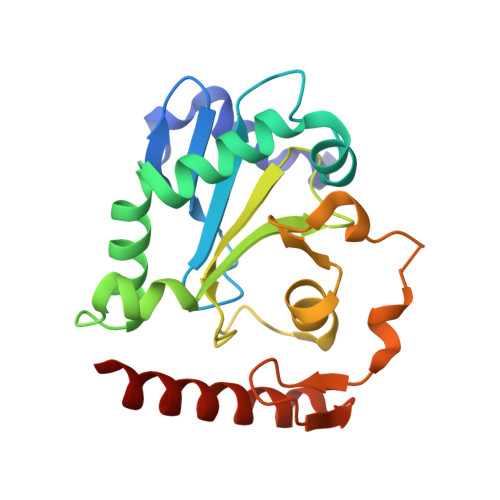
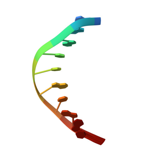
DNA3'pp5'G de-capping activity of aprataxin: effect of cap nucleoside analogs and structural basis for guanosine recognition.

Chauleau, M.Jacewicz, A.Shuman, S.

(2015) Nucleic Acids Res 43: 6075-6083

  • DOI: https://doi.org/10.1093/nar/gkv501
  • Primary Citation Related Structures: 
    4XBA, 4YKL

  • PubMed Abstract: 

    DNA3'pp5'G caps synthesized by the 3'-PO4/5'-OH ligase RtcB have a strong impact on enzymatic reactions at DNA 3'-OH ends. Aprataxin, an enzyme that repairs A5'pp5'DNA ends formed during abortive ligation by classic 3'-OH/5'-PO4 ligases, is also a DNA 3' de-capping enzyme, converting DNAppG to DNA3'p and GMP. By taking advantage of RtcB's ability to utilize certain GTP analogs to synthesize DNAppN caps, we show that aprataxin hydrolyzes inosine and 6-O-methylguanosine caps, but is not adept at removing a deoxyguanosine cap. We report a 1.5 Å crystal structure of aprataxin in a complex with GMP, which reveals that: (i) GMP binds at the same position and in the same anti nucleoside conformation as AMP; and (ii) aprataxin makes more extensive nucleobase contacts with guanine than with adenine, via a hydrogen bonding network to the guanine O6, N1, N2 base edge. Alanine mutations of catalytic residues His147 and His149 abolish DNAppG de-capping activity, suggesting that the 3' de-guanylylation and 5' de-adenylylation reactions follow the same pathway of nucleotidyl transfer through a covalent aprataxin-(His147)-NMP intermediate. Alanine mutation of Asp63, which coordinates the guanosine ribose hydroxyls, impairs DNAppG de-capping.


  • Organizational Affiliation
    • Molecular Biology Program, Sloan-Kettering Institute, New York, NY 10065, USA.

Macromolecule Content 

  • Total Structure Weight: 27.05 kDa 
  • Atom Count: 1,898 
  • Modeled Residue Count: 205 
  • Deposited Residue Count: 210 
  • Unique protein chains: 1
  • Unique nucleic acid chains: 1

Macromolecules


Find similar proteins by:|  3D Structure
Entity ID: 1
MoleculeChains  Sequence LengthOrganismDetailsImage
Aprataxin-like proteinA [auth B]200Schizosaccharomyces pombe 972h-Mutation(s): 0 
Gene Names: hnt3SPCC18.09c
EC: 3 (PDB Primary Data), 3.6.1.72 (UniProt), 3.6.1.71 (UniProt)
UniProt
Find proteins for O74859 (Schizosaccharomyces pombe (strain 972 / ATCC 24843))
Explore O74859 
Go to UniProtKB:  O74859
Entity Groups
Sequence Clusters30% Identity50% Identity70% Identity90% Identity95% Identity100% Identity
UniProt GroupO74859
Sequence Annotations
Expand
Reference Sequence
Find similar nucleic acids by:  Sequence
Entity ID: 2
MoleculeChains LengthOrganismImage
DNA (5'-D(*GP*AP*AP*TP*CP*AP*TP*AP*AP*C)-3')B [auth A]10synthetic construct
Sequence Annotations
Expand
Reference Sequence

Small Molecules

Ligands 4 Unique
IDChains Name / Formula / InChI Key2D Diagram3D Interactions
GMP

Query on GMP


Download:Ideal Coordinates CCD File
D [auth B]GUANOSINE
C10 H13 N5 O5
NYHBQMYGNKIUIF-UUOKFMHZSA-N
GOL

Query on GOL


Download:Ideal Coordinates CCD File
E [auth B],
F [auth B],
G [auth B],
H [auth B]
GLYCEROL
C3 H8 O3
PEDCQBHIVMGVHV-UHFFFAOYSA-N
ZN

Query on ZN


Download:Ideal Coordinates CCD File
C [auth B]ZINC ION
Zn
PTFCDOFLOPIGGS-UHFFFAOYSA-N
CL

Query on CL


Download:Ideal Coordinates CCD File
I [auth B],
J [auth B]
CHLORIDE ION
Cl
VEXZGXHMUGYJMC-UHFFFAOYSA-M

Experimental Data & Validation

Experimental Data

  • Method: X-RAY DIFFRACTION
  • Resolution: 2.25 Å
  • R-Value Free:  0.240 (Depositor), 0.246 (DCC) 
  • R-Value Work:  0.180 (Depositor), 0.186 (DCC) 
  • R-Value Observed: 0.184 (Depositor) 
Space Group: P 3 2 1
Unit Cell:
Length ( Å )Angle ( ˚ )
a = 119.083α = 90
b = 119.083β = 90
c = 36.564γ = 120
Software Package:
Software NamePurpose
PHENIXrefinement
XDSdata reduction
Aimlessdata scaling
PHASERphasing

Structure Validation

View Full Validation Report


Ligand Structure Quality Assessment 


Entry History 

& Funding Information

Deposition Data


Funding OrganizationLocationGrant Number
National Institutes of Health/National Institute of General Medical Sciences (NIH/NIGMS)United StatesGM46330

Revision History  (Full details and data files)

  • Version 1.0: 2015-06-03
    Type: Initial release
  • Version 1.1: 2015-06-10
    Changes: Database references
  • Version 1.2: 2015-07-22
    Changes: Database references
  • Version 1.3: 2016-07-27
    Changes: Data collection
  • Version 1.4: 2017-09-13
    Changes: Author supporting evidence, Derived calculations
  • Version 1.5: 2019-12-25
    Changes: Author supporting evidence
  • Version 1.6: 2023-09-27
    Changes: Data collection, Database references, Refinement description