4YKB

Structure of GUN4 from Chlamydomonas reinhardtii


Experimental Data Snapshot

  • Method: X-RAY DIFFRACTION
  • Resolution: 3.50 Å
  • R-Value Free: 0.313 
  • R-Value Work: 0.256 
  • R-Value Observed: 0.259 

wwPDB Validation   3D Report Full Report


This is version 1.2 of the entry. See complete history


Literature

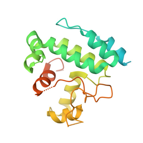
Structure of GUN4 from Chlamydomonas reinhardtii.

Tarahi Tabrizi, S.Langley, D.B.Harrop, S.J.Duff, A.P.Willows, R.D.

(2015) Acta Crystallogr F Struct Biol Commun 71: 1094-1099

  • DOI: https://doi.org/10.1107/S2053230X15012248
  • Primary Citation of Related Structures:  
    4YKB

  • PubMed Abstract: 

    The genomes uncoupled 4 (GUN4) protein stimulates chlorophyll biosynthesis by increasing the activity of Mg-chelatase, the enzyme that inserts magnesium into protoporphyrin IX (PPIX) in the chlorophyll biosynthesis pathway. One of the roles of GUN4 is in binding PPIX and Mg-PPIX. In eukaryotes, GUN4 also participates in plastid-to-nucleus signalling, although the mechanism for this is unclear. Here, the first crystal structure of a eukaryotic GUN4, from Chlamydomonas reinhardtii, is presented. The structure is in broad agreement with those of previously solved cyanobacterial structures. Most interestingly, conformational divergence is restricted to several loops which cover the porphyrin-binding cleft. The conformational dynamics suggested by this ensemble of structures lend support to the understanding of how GUN4 binds PPIX or Mg-PPIX.


  • Organizational Affiliation

    Department of Chemistry and Biomolecular Sciences, Macquarie University, Sydney, NSW 2109, Australia.


Macromolecules
Find similar proteins by:  (by identity cutoff)  |  3D Structure
Entity ID: 1
MoleculeChains Sequence LengthOrganismDetailsImage
Tetrapyrrole-binding protein
A, B, C, D, E
A, B, C, D, E, F
229Chlamydomonas reinhardtiiMutation(s): 0 
Gene Names: GUN4CHLREDRAFT_205768
UniProt
Find proteins for A8I5N5 (Chlamydomonas reinhardtii)
Explore A8I5N5 
Go to UniProtKB:  A8I5N5
Entity Groups  
Sequence Clusters30% Identity50% Identity70% Identity90% Identity95% Identity100% Identity
UniProt GroupA8I5N5
Sequence Annotations
Expand
  • Reference Sequence
Experimental Data & Validation

Experimental Data

  • Method: X-RAY DIFFRACTION
  • Resolution: 3.50 Å
  • R-Value Free: 0.313 
  • R-Value Work: 0.256 
  • R-Value Observed: 0.259 
  • Space Group: P 32 2 1
Unit Cell:
Length ( Å )Angle ( ˚ )
a = 114.998α = 90
b = 114.998β = 90
c = 141.193γ = 120
Software Package:
Software NamePurpose
REFMACrefinement
MOSFLMdata reduction
Aimlessdata scaling
PHASERphasing

Structure Validation

View Full Validation Report


Entry History 

Deposition Data

Revision History  (Full details and data files)

  • Version 1.0: 2015-08-12
    Type: Initial release
  • Version 1.1: 2018-05-16
    Changes: Data collection, Database references, Derived calculations
  • Version 1.2: 2023-09-27
    Changes: Data collection, Database references, Refinement description