


Structure-based design of a bisphosphonate 5'(3')-deoxyribonucleotidase inhibitor
Pachl, P., Simak, O., Rezacova, P., Fabry, M., Budesinsky, M., Rosenberg, I., Brynda, J.(2015) Medchemcomm 6: 1635-1638
Experimental Data Snapshot
Starting Model: experimental
View more details
(2015) Medchemcomm 6: 1635-1638
Entity ID: 1 | |||||
|---|---|---|---|---|---|
| Molecule | Chains | Sequence Length | Organism | Details | Image |
| 5'(3')-deoxyribonucleotidase, mitochondrial | 201 | Homo sapiens | Mutation(s): 0 Gene Names: NT5M, DNT2 EC: 3.1.3 | 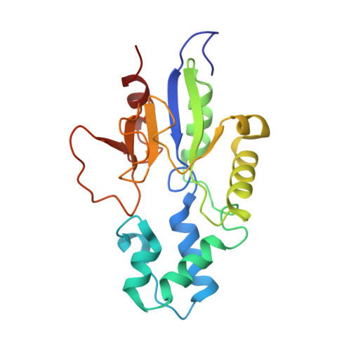 | |
UniProt & NIH Common Fund Data Resources | |||||
PHAROS: Q9NPB1 GTEx: ENSG00000205309 | |||||
Entity Groups | |||||
| Sequence Clusters | 30% Identity50% Identity70% Identity90% Identity95% Identity100% Identity | ||||
| UniProt Group | Q9NPB1 | ||||
Sequence AnnotationsExpand | |||||
Reference Sequence | |||||
| Ligands 6 Unique | |||||
|---|---|---|---|---|---|
| ID | Chains | Name / Formula / InChI Key | 2D Diagram | 3D Interactions | |
| 2O2 Download:Ideal Coordinates CCD File | G [auth A] | 1-{2-deoxy-3,5-O-[phenyl(phosphono)methylidene]-beta-D-threo-pentofuranosyl}-5-[(E)-2-phosphonoethenyl]pyrimidine-2,4(1H,3H)-dione C18 H20 N2 O11 P2 AYSYVLQGVXZPIY-OAIWFRFLSA-N |  | ||
| TRS Download:Ideal Coordinates CCD File | H [auth A] | 2-AMINO-2-HYDROXYMETHYL-PROPANE-1,3-DIOL C4 H12 N O3 LENZDBCJOHFCAS-UHFFFAOYSA-O |  | ||
| PEG Download:Ideal Coordinates CCD File | I [auth A], J [auth A], K [auth A], L [auth A], M [auth A] | DI(HYDROXYETHYL)ETHER C4 H10 O3 MTHSVFCYNBDYFN-UHFFFAOYSA-N |  | ||
| PO4 Download:Ideal Coordinates CCD File | N [auth A] | PHOSPHATE ION O4 P NBIIXXVUZAFLBC-UHFFFAOYSA-K |  | ||
| GOL Download:Ideal Coordinates CCD File | C [auth A], D [auth A], E [auth A], F [auth A] | GLYCEROL C3 H8 O3 PEDCQBHIVMGVHV-UHFFFAOYSA-N |  | ||
| MG Download:Ideal Coordinates CCD File | B [auth A] | MAGNESIUM ION Mg JLVVSXFLKOJNIY-UHFFFAOYSA-N |  | ||
| Length ( Å ) | Angle ( ˚ ) |
|---|---|
| a = 73.87 | α = 90 |
| b = 73.87 | β = 90 |
| c = 106.19 | γ = 90 |
| Software Name | Purpose |
|---|---|
| PDB_EXTRACT | data extraction |
| SCALA | data scaling |
| REFMAC | refinement |
| MOSFLM | data reduction |
| REFMAC | phasing |
| Funding Organization | Location | Grant Number |
|---|---|---|
| Grant Agency of the Czech Republic | Czech Republic | -- |


