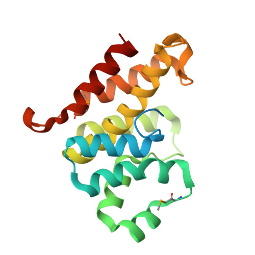
1.85 angstrom crystal structure of lmo0812 from Listeria monocytogenes EGD-e
Krishna, S.N., Light, S.H., Filippova, E.V., Minasov, G., Kiryukhina, O., Jedrzejczak, R., Joachimiak, A., Anderson, W.F., Midwest Center for Structural Genomics (MCSG)To be published.