


A cytochrome c-calixarene structure
Mallon, M.M., McGovern, R.E., McCarty, A.A., Crowley, P.B.To be published.
Experimental Data Snapshot
Starting Model: experimental
View more details
Entity ID: 1 | |||||
|---|---|---|---|---|---|
| Molecule | Chains | Sequence Length | Organism | Details | Image |
| Cytochrome c iso-1 | 108 | Saccharomyces cerevisiae S288C | Mutation(s): 1 Gene Names: CYC1, YJR048W, J1653 | 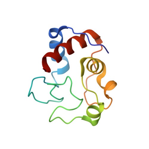 | |
UniProt | |||||
Entity Groups | |||||
| Sequence Clusters | 30% Identity50% Identity70% Identity90% Identity95% Identity100% Identity | ||||
| UniProt Group | P00044 | ||||
Sequence AnnotationsExpand | |||||
Reference Sequence | |||||
| Ligands 3 Unique | |||||
|---|---|---|---|---|---|
| ID | Chains | Name / Formula / InChI Key | 2D Diagram | 3D Interactions | |
| T3Y Download:Ideal Coordinates CCD File | D [auth A], F [auth B], G [auth B] | 25,26,27,28-tetrahydroxypentacyclo[19.3.1.1~3,7~.1~9,13~.1~15,19~]octacosa-1(25),3(28),4,6,9(27),10,12,15(26),16,18,21,23-dodecaene-5,11,17,23-tetrasulfonic acid C28 H24 O16 S4 JFYBCAFLVNKHHG-UHFFFAOYSA-N |  | ||
| HEC Download:Ideal Coordinates CCD File | C [auth A], E [auth B] | HEME C C34 H34 Fe N4 O4 HXQIYSLZKNYNMH-LJNAALQVSA-N |  | ||
| GOL Download:Ideal Coordinates CCD File | H [auth B] | GLYCEROL C3 H8 O3 PEDCQBHIVMGVHV-UHFFFAOYSA-N |  | ||
| Length ( Å ) | Angle ( ˚ ) |
|---|---|
| a = 35.85 | α = 90 |
| b = 56.07 | β = 90 |
| c = 108.04 | γ = 90 |
| Software Name | Purpose |
|---|---|
| REFMAC | refinement |
| MOSFLM | data reduction |
| SCALA | data scaling |
| PHASER | phasing |
| PDB_EXTRACT | data extraction |


