4YD1 | pdb_00004yd1

Ternary complex of human DNA Polymerase Mu with 2-nt gapped DNA substrate and an incoming nonhydrolyzable dUMPNPP


Experimental Data Snapshot

  • Method: X-RAY DIFFRACTION
  • Resolution: 1.75 Å
  • R-Value Free: 
    0.190 (Depositor), 0.190 (DCC) 
  • R-Value Work: 
    0.163 (Depositor), 0.164 (DCC) 
  • R-Value Observed: 
    0.164 (Depositor) 

Starting Model: experimental
View more details

wwPDB Validation 3D Report Full Report

Validation slider image for 4YD1

Ligand Structure Quality Assessment 


This is version 1.5 of the entry. See complete history

Literature

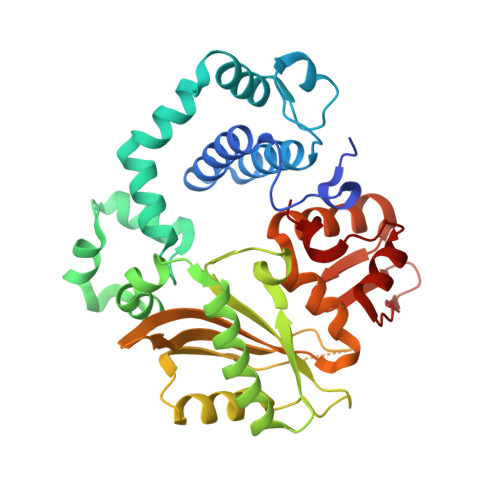
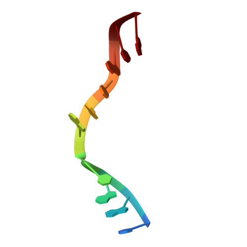
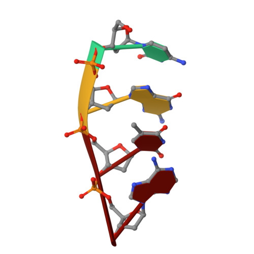
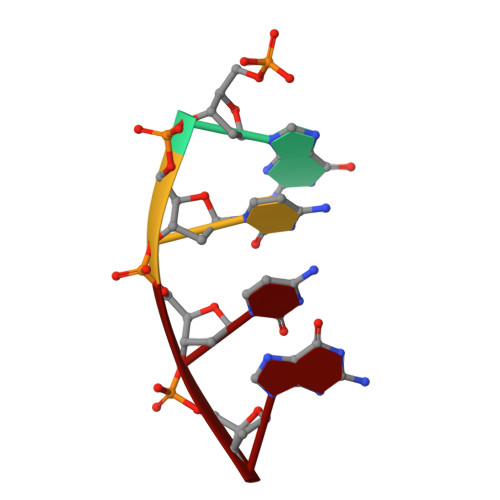
Creative template-dependent synthesis by human polymerase mu.

Moon, A.F.Gosavi, R.A.Kunkel, T.A.Pedersen, L.C.Bebenek, K.

(2015) Proc Natl Acad Sci U S A 112: E4530-E4536

  • DOI: https://doi.org/10.1073/pnas.1505798112
  • Primary Citation Related Structures: 
    4YCX, 4YD1, 4YD2

  • PubMed Abstract: 

    Among the many proteins used to repair DNA double-strand breaks by nonhomologous end joining (NHEJ) are two related family X DNA polymerases, Pol λ and Pol µ. Which of these two polymerases is preferentially used for filling DNA gaps during NHEJ partly depends on sequence complementarity at the break, with Pol λ and Pol µ repairing complementary and noncomplementary ends, respectively. To better understand these substrate preferences, we present crystal structures of Pol µ on a 2-nt gapped DNA substrate, representing three steps of the catalytic cycle. In striking contrast to Pol λ, Pol µ "skips" the first available template nucleotide, instead using the template base at the 5' end of the gap to direct nucleotide binding and incorporation. This remarkable divergence from canonical 3'-end gap filling is consistent with data on end-joining substrate specificity in cells, and provides insights into polymerase substrate choices during NHEJ.


  • Organizational Affiliation
    • Genome Integrity and Structural Biology Laboratory, National Institute of Environmental Health Sciences, National Institutes of Health, Research Triangle Park, NC 27709.

Macromolecule Content 

  • Total Structure Weight: 46.29 kDa 
  • Atom Count: 3,581 
  • Modeled Residue Count: 352 
  • Deposited Residue Count: 372 
  • Unique protein chains: 1
  • Unique nucleic acid chains: 3

Macromolecules


Find similar proteins by:|  3D Structure
Entity ID: 1
MoleculeChains  Sequence LengthOrganismDetailsImage
DNA-directed DNA/RNA polymerase mu354Homo sapiensMutation(s): 1 
Gene Names: POLMpolmu
EC: 2.7.7.7
UniProt & NIH Common Fund Data Resources
Find proteins for Q9NP87 (Homo sapiens)
Explore Q9NP87 
Go to UniProtKB:  Q9NP87
PHAROS:  Q9NP87
GTEx:  ENSG00000122678 
Entity Groups
Sequence Clusters30% Identity50% Identity70% Identity90% Identity95% Identity100% Identity
UniProt GroupQ9NP87
Sequence Annotations
Expand
Reference Sequence
Find similar nucleic acids by:  Sequence
Entity ID: 2
MoleculeChains LengthOrganismImage
DNA (5'-D(*CP*GP*GP*CP*AP*AP*TP*AP*CP*G)-3')B [auth T]10Homo sapiens
Sequence Annotations
Expand
Reference Sequence
Find similar nucleic acids by:  Sequence
Entity ID: 3
MoleculeChains LengthOrganismImage
DNA (5'-D(*CP*GP*TP*A)-3')C [auth P]4Homo sapiens
Sequence Annotations
Expand
Reference Sequence
Find similar nucleic acids by:  Sequence
Entity ID: 4
MoleculeChains LengthOrganismImage
DNA (5'-D(P*GP*CP*CP*G)-3')4Homo sapiens
Sequence Annotations
Expand
Reference Sequence

Small Molecules

Ligands 6 Unique
IDChains Name / Formula / InChI Key2D Diagram3D Interactions
DUP

Query on DUP


Download:Ideal Coordinates CCD File
K [auth A]2'-DEOXYURIDINE 5'-ALPHA,BETA-IMIDO-TRIPHOSPHATE
C9 H16 N3 O13 P3
XZLLMTSKYYYJLH-SHYZEUOFSA-N
GOL

Query on GOL


Download:Ideal Coordinates CCD File
H [auth A],
I [auth A]
GLYCEROL
C3 H8 O3
PEDCQBHIVMGVHV-UHFFFAOYSA-N
EDO

Query on EDO


Download:Ideal Coordinates CCD File
G [auth A],
J [auth A]
1,2-ETHANEDIOL
C2 H6 O2
LYCAIKOWRPUZTN-UHFFFAOYSA-N
CL

Query on CL


Download:Ideal Coordinates CCD File
L [auth A],
M [auth A],
N [auth A],
O [auth A]
CHLORIDE ION
Cl
VEXZGXHMUGYJMC-UHFFFAOYSA-M
MG

Query on MG


Download:Ideal Coordinates CCD File
E [auth A],
F [auth A],
Q [auth T]
MAGNESIUM ION
Mg
JLVVSXFLKOJNIY-UHFFFAOYSA-N
NA

Query on NA


Download:Ideal Coordinates CCD File
P [auth A]SODIUM ION
Na
FKNQFGJONOIPTF-UHFFFAOYSA-N

Experimental Data & Validation

Experimental Data

  • Method: X-RAY DIFFRACTION
  • Resolution: 1.75 Å
  • R-Value Free:  0.190 (Depositor), 0.190 (DCC) 
  • R-Value Work:  0.163 (Depositor), 0.164 (DCC) 
  • R-Value Observed: 0.164 (Depositor) 
Space Group: P 21 21 21
Unit Cell:
Length ( Å )Angle ( ˚ )
a = 60.259α = 90
b = 69.042β = 90
c = 116.988γ = 90
Software Package:
Software NamePurpose
PHENIXrefinement
HKL-2000data reduction
HKL-2000data scaling
Cootmodel building
PHASERphasing

Structure Validation

View Full Validation Report


Ligand Structure Quality Assessment 


Entry History 

& Funding Information

Deposition Data


Funding OrganizationLocationGrant Number
National Institutes of Health/National Institute of Environmental Health Sciences (NIH/NIEHS)United States1ZIA ES102645-03
National Institutes of Health/National Institute of Environmental Health Sciences (NIH/NIEHS)United StatesZ01 ES065070

Revision History  (Full details and data files)

  • Version 1.0: 2015-08-05
    Type: Initial release
  • Version 1.1: 2015-08-19
    Changes: Database references
  • Version 1.2: 2015-08-26
    Changes: Database references
  • Version 1.3: 2017-09-20
    Changes: Author supporting evidence, Database references, Derived calculations
  • Version 1.4: 2019-12-18
    Changes: Author supporting evidence
  • Version 1.5: 2023-09-27
    Changes: Data collection, Database references, Derived calculations, Refinement description