4YCE | pdb_00004yce

Crystal structure of the R111K:Y134F:T54V:R132Q:P39Q:R59Y mutant of human cellular retinoic acid binding proteinii with retinal at 1.95 angstrom- visible light irradiated crystal for 1 hour - 2nd cycle


Experimental Data Snapshot

  • Method: X-RAY DIFFRACTION
  • Resolution: 1.95 Å
  • R-Value Free: 
    0.238 (Depositor), 0.237 (DCC) 
  • R-Value Work: 
    0.190 (Depositor), 0.188 (DCC) 
  • R-Value Observed: 
    0.192 (Depositor) 

Starting Model: experimental
View more details

wwPDB Validation 3D Report Full Report

Validation slider image for 4YCE

Ligand Structure Quality Assessment 


This is version 1.5 of the entry. See complete history

Literature

A Photoisomerizing Rhodopsin Mimic Observed at Atomic Resolution.

Nosrati, M.Berbasova, T.Vasileiou, C.Borhan, B.Geiger, J.H.

(2016) J Am Chem Soc 138: 8802-8808

  • DOI: https://doi.org/10.1021/jacs.6b03681
  • Primary Citation Related Structures: 
    4YBP, 4YBU, 4YCE, 4YCH, 4YDA, 4YDB, 4YFP, 4YFQ, 4YFR, 4YGG, 4YGH, 4YGZ, 4YH0, 4YKM, 4YKO

  • PubMed Abstract: 

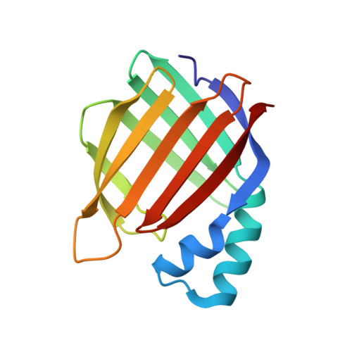
    The members of the rhodopsin family of proteins are involved in many essential light-dependent processes in biology. Specific photoisomerization of the protein-bound retinylidene PSB at a specified wavelength range of light is at the heart of all of these systems. Nonetheless, it has been difficult to reproduce in an engineered system. We have developed rhodopsin mimics, using intracellular lipid binding protein family members as scaffolds, to study fundamental aspects of protein/chromophore interactions. Herein we describe a system that specifically isomerizes the retinylidene protonated Schiff base both thermally and photochemically. This isomerization has been characterized at atomic resolution by quantitatively interconverting the isomers in the crystal both thermally and photochemically. This event is accompanied by a large pKa change of the imine similar to the pKa changes observed in bacteriorhodopsin and visual opsins during isomerization.


  • Organizational Affiliation
    • Department of Chemistry, Michigan State University , East Lansing, Michigan 48824, United States.

Macromolecule Content 

  • Total Structure Weight: 15.83 kDa 
  • Atom Count: 1,297 
  • Modeled Residue Count: 137 
  • Deposited Residue Count: 137 
  • Unique protein chains: 1

Macromolecules

Find similar proteins by:|  3D Structure
Entity ID: 1
MoleculeChains  Sequence LengthOrganismDetailsImage
Cellular retinoic acid-binding protein 2137Homo sapiensMutation(s): 6 
Gene Names: CRABP2
UniProt & NIH Common Fund Data Resources
Find proteins for P29373 (Homo sapiens)
Explore P29373 
Go to UniProtKB:  P29373
PHAROS:  P29373
GTEx:  ENSG00000143320 
Entity Groups
Sequence Clusters30% Identity50% Identity70% Identity90% Identity95% Identity100% Identity
UniProt GroupP29373
Sequence Annotations
Expand
Reference Sequence

Small Molecules

Ligands 1 Unique
IDChains Name / Formula / InChI Key2D Diagram3D Interactions
RET

Query on RET


Download:Ideal Coordinates CCD File
B [auth A]RETINAL
C20 H28 O
NCYCYZXNIZJOKI-OVSJKPMPSA-N

Experimental Data & Validation

Experimental Data

  • Method: X-RAY DIFFRACTION
  • Resolution: 1.95 Å
  • R-Value Free:  0.238 (Depositor), 0.237 (DCC) 
  • R-Value Work:  0.190 (Depositor), 0.188 (DCC) 
  • R-Value Observed: 0.192 (Depositor) 
Space Group: P 31 2 1
Unit Cell:
Length ( Å )Angle ( ˚ )
a = 58.599α = 90
b = 58.599β = 90
c = 99.854γ = 120
Software Package:
Software NamePurpose
PHENIXrefinement
HKL-2000data reduction
HKL-2000data scaling
MOLREPphasing

Structure Validation

View Full Validation Report


Ligand Structure Quality Assessment 


Entry History 

Deposition Data

Revision History  (Full details and data files)

  • Version 1.0: 2016-02-24
    Type: Initial release
  • Version 1.1: 2016-06-15
    Changes: Structure summary
  • Version 1.2: 2016-06-29
    Changes: Database references
  • Version 1.3: 2016-08-03
    Changes: Database references
  • Version 1.4: 2023-09-27
    Changes: Data collection, Database references, Derived calculations, Refinement description
  • Version 1.5: 2024-11-13
    Changes: Structure summary