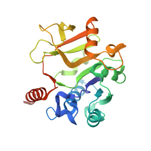
4Y7U | pdb_00004y7u

Structural analysis of MurU


Experimental Data Snapshot

  • Method: X-RAY DIFFRACTION
  • Resolution: 1.70 Å
  • R-Value Free: 
    0.264 (Depositor), 0.260 (DCC) 
  • R-Value Work: 
    0.228 (Depositor), 0.230 (DCC) 
  • R-Value Observed: 
    0.230 (Depositor) 

wwPDB Validation   3D Report Full Report


Ligand Structure Quality Assessment 

Created with Raphaël 2.3.0Worse 01 BetterLigand structure goodness of fit to experimental dataBest fitted 2KHClick on this verticalbar to view detailsBest fitted 491Click on this verticalbar to view details

This is version 2.1 of the entry. See complete history


Literature

Crystal Structure of the N-Acetylmuramic Acid alpha-1-Phosphate (MurNAc-alpha 1-P) Uridylyltransferase MurU, a Minimal Sugar Nucleotidyltransferase and Potential Drug Target Enzyme in Gram-negative Pathogens.

Renner-Schneck, M.Hinderberger, I.Gisin, J.Exner, T.Mayer, C.Stehle, T.

(2015) J Biological Chem 290: 10804-10813

  • DOI: https://doi.org/10.1074/jbc.M114.620989
  • Primary Citation of Related Structures:  
    4Y7T, 4Y7U, 4Y7V

  • PubMed Abstract: 

    The N-acetylmuramic acid α-1-phosphate (MurNAc-α1-P) uridylyltransferase MurU catalyzes the synthesis of uridine diphosphate (UDP)-MurNAc, a crucial precursor of the bacterial peptidoglycan cell wall. MurU is part of a recently identified cell wall recycling pathway in Gram-negative bacteria that bypasses the general de novo biosynthesis of UDP-MurNAc and contributes to high intrinsic resistance to the antibiotic fosfomycin, which targets UDP-MurNAc de novo biosynthesis. To provide insights into substrate binding and specificity, we solved crystal structures of MurU of Pseudomonas putida in native and ligand-bound states at high resolution. With the help of these structures, critical enzyme-substrate interactions were identified that enable tight binding of MurNAc-α1-P to the active site of MurU. The MurU structures define a "minimal domain" required for general nucleotidyltransferase activity. They furthermore provide a structural basis for the chemical design of inhibitors of MurU that could serve as novel drugs in combination therapy against multidrug-resistant Gram-negative pathogens.


  • Organizational Affiliation

    From the Interfaculty Institute of Biochemistry (IFIB).


Macromolecules
Find similar proteins by:  (by identity cutoff)  |  3D Structure
Entity ID: 1
MoleculeChains Sequence LengthOrganismDetailsImage
Nucleotidyl transferase231Pseudomonas putida BIRD-1Mutation(s): 0 
Gene Names: PPUBIRD1_0444
EC: 2.7.7.99
UniProt
Find proteins for Q88QT2 (Pseudomonas putida (strain ATCC 47054 / DSM 6125 / CFBP 8728 / NCIMB 11950 / KT2440))
Explore Q88QT2 
Go to UniProtKB:  Q88QT2
Entity Groups  
Sequence Clusters30% Identity50% Identity70% Identity90% Identity95% Identity100% Identity
UniProt GroupQ88QT2
Sequence Annotations
Expand
  • Reference Sequence
Small Molecules
Ligands 5 Unique
IDChains Name / Formula / InChI Key2D Diagram3D Interactions
2KH
Query on 2KH

Download Ideal Coordinates CCD File 
G [auth A]5'-O-[(S)-hydroxy{[(S)-hydroxy(phosphonooxy)phosphoryl]amino}phosphoryl]uridine
C9 H16 N3 O14 P3
OZIBFYOFLVBDIY-XVFCMESISA-N
491
Query on 491

Download Ideal Coordinates CCD File 
H [auth A]2-acetamido-3-O-[(1R)-1-carboxyethyl]-2-deoxy-1-O-phosphono-alpha-D-glucopyranose
C11 H20 N O11 P
DTSXRQWOCZUNPL-YVNCZSHWSA-N
SO4
Query on SO4

Download Ideal Coordinates CCD File 
D [auth A]SULFATE ION
O4 S
QAOWNCQODCNURD-UHFFFAOYSA-L
GOL
Query on GOL

Download Ideal Coordinates CCD File 
B [auth A],
C [auth A]
GLYCEROL
C3 H8 O3
PEDCQBHIVMGVHV-UHFFFAOYSA-N
MG
Query on MG

Download Ideal Coordinates CCD File 
E [auth A],
F [auth A]
MAGNESIUM ION
Mg
JLVVSXFLKOJNIY-UHFFFAOYSA-N
Experimental Data & Validation

Experimental Data

  • Method: X-RAY DIFFRACTION
  • Resolution: 1.70 Å
  • R-Value Free:  0.264 (Depositor), 0.260 (DCC) 
  • R-Value Work:  0.228 (Depositor), 0.230 (DCC) 
  • R-Value Observed: 0.230 (Depositor) 
Space Group: P 61 2 2
Unit Cell:
Length ( Å )Angle ( ˚ )
a = 72.75α = 90
b = 72.75β = 90
c = 163.13γ = 120
Software Package:
Software NamePurpose
XDSdata reduction
PHENIXrefinement
PDB_EXTRACTdata extraction

Structure Validation

View Full Validation Report


Ligand Structure Quality Assessment 

Created with Raphaël 2.3.0Worse 01 BetterLigand structure goodness of fit to experimental dataBest fitted 2KHClick on this verticalbar to view detailsBest fitted 491Click on this verticalbar to view details

Entry History 

Deposition Data

Revision History  (Full details and data files)

  • Version 1.0: 2015-03-18
    Type: Initial release
  • Version 1.1: 2015-03-25
    Changes: Database references
  • Version 1.2: 2015-05-06
    Changes: Database references
  • Version 2.0: 2020-07-29
    Type: Remediation
    Reason: Carbohydrate remediation
    Changes: Atomic model, Derived calculations, Structure summary
  • Version 2.1: 2024-05-08
    Changes: Data collection, Database references, Structure summary