4Y14 | pdb_00004y14

Structure of protein tyrosine phosphatase 1B complexed with inhibitor (PTP1B:CPT157633)


Experimental Data Snapshot

  • Method: X-RAY DIFFRACTION
  • Resolution: 1.90 Å
  • R-Value Free: 
    0.206 (Depositor), 0.208 (DCC) 
  • R-Value Work: 
    0.166 (Depositor), 0.169 (DCC) 
  • R-Value Observed: 
    0.168 (Depositor) 

Starting Model: experimental
View more details

wwPDB Validation 3D Report Full Report

Validation slider image for 4Y14

Ligand Structure Quality Assessment 


This is version 1.2 of the entry. See complete history

Literature

PTP1B inhibition suggests a therapeutic strategy for Rett syndrome.

Krishnan, N.Krishnan, K.Connors, C.R.Choy, M.S.Page, R.Peti, W.Van Aelst, L.Shea, S.D.Tonks, N.K.

(2015) J Clin Invest 125: 3163-3177

  • DOI: https://doi.org/10.1172/JCI80323
  • Primary Citation Related Structures: 
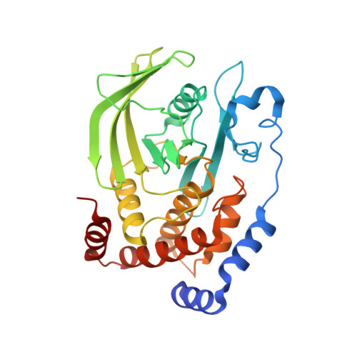
    4Y14

  • PubMed Abstract: 

    The X-linked neurological disorder Rett syndrome (RTT) presents with autistic features and is caused primarily by mutations in a transcriptional regulator, methyl CpG-binding protein 2 (MECP2). Current treatment options for RTT are limited to alleviating some neurological symptoms; hence, more effective therapeutic strategies are needed. We identified the protein tyrosine phosphatase PTP1B as a therapeutic candidate for treatment of RTT. We demonstrated that the PTPN1 gene, which encodes PTP1B, was a target of MECP2 and that disruption of MECP2 function was associated with increased levels of PTP1B in RTT models. Pharmacological inhibition of PTP1B ameliorated the effects of MECP2 disruption in mouse models of RTT, including improved survival in young male (Mecp2-/y) mice and improved behavior in female heterozygous (Mecp2-/+) mice. We demonstrated that PTP1B was a negative regulator of tyrosine phosphorylation of the tyrosine kinase TRKB, the receptor for brain-derived neurotrophic factor (BDNF). Therefore, the elevated PTP1B that accompanies disruption of MECP2 function in RTT represents a barrier to BDNF signaling. Inhibition of PTP1B led to increased tyrosine phosphorylation of TRKB in the brain, which would augment BDNF signaling. This study presents PTP1B as a mechanism-based therapeutic target for RTT, validating a unique strategy for treating the disease by modifying signal transduction pathways with small-molecule drugs.

Macromolecule Content 

  • Total Structure Weight: 73.16 kDa 
  • Atom Count: 5,375 
  • Modeled Residue Count: 589 
  • Deposited Residue Count: 616 
  • Unique protein chains: 1

Macromolecules

Find similar proteins by:|  3D Structure
Entity ID: 1
MoleculeChains  Sequence LengthOrganismDetailsImage
Tyrosine-protein phosphatase non-receptor type 1
A, B
308Homo sapiensMutation(s): 0 
Gene Names: PTPN1PTP1B
EC: 3.1.3.48
UniProt & NIH Common Fund Data Resources
Find proteins for P18031 (Homo sapiens)
Explore P18031 
Go to UniProtKB:  P18031
PHAROS:  P18031
GTEx:  ENSG00000196396 
Entity Groups
Sequence Clusters30% Identity50% Identity70% Identity90% Identity95% Identity100% Identity
UniProt GroupP18031
Sequence Annotations
Expand
Reference Sequence

Small Molecules

Ligands 4 Unique
IDChains Name / Formula / InChI Key2D Diagram3D Interactions
C0A

Query on C0A


Download:Ideal Coordinates CCD File
I [auth A],
M [auth B]
3-bromo-4-[difluoro(phosphono)methyl]-N-methyl-Nalpha-(methylsulfonyl)-L-phenylalaninamide
C12 H16 Br F2 N2 O6 P S
GNQQLTLSVIWPPQ-JTQLQIEISA-N
TRS

Query on TRS


Download:Ideal Coordinates CCD File
F [auth A],
G [auth A],
L [auth B]
2-AMINO-2-HYDROXYMETHYL-PROPANE-1,3-DIOL
C4 H12 N O3
LENZDBCJOHFCAS-UHFFFAOYSA-O
GOL

Query on GOL


Download:Ideal Coordinates CCD File
H [auth A]GLYCEROL
C3 H8 O3
PEDCQBHIVMGVHV-UHFFFAOYSA-N
CL

Query on CL


Download:Ideal Coordinates CCD File
C [auth A],
D [auth A],
E [auth A],
J [auth B],
K [auth B]
CHLORIDE ION
Cl
VEXZGXHMUGYJMC-UHFFFAOYSA-M

Experimental Data & Validation

Experimental Data

  • Method: X-RAY DIFFRACTION
  • Resolution: 1.90 Å
  • R-Value Free:  0.206 (Depositor), 0.208 (DCC) 
  • R-Value Work:  0.166 (Depositor), 0.169 (DCC) 
  • R-Value Observed: 0.168 (Depositor) 
Space Group: P 1 21 1
Unit Cell:
Length ( Å )Angle ( ˚ )
a = 52.167α = 90
b = 71.748β = 94.3
c = 88.01γ = 90
Software Package:
Software NamePurpose
PHENIXrefinement
HKL-3000data reduction
PHASERphasing
PDB_EXTRACTdata extraction
PHENIXmodel building
HKL-3000data scaling

Structure Validation

View Full Validation Report


Ligand Structure Quality Assessment 


Entry History 

Deposition Data

Revision History  (Full details and data files)

  • Version 1.0: 2015-08-05
    Type: Initial release
  • Version 1.1: 2015-08-26
    Changes: Database references
  • Version 1.2: 2023-09-27
    Changes: Data collection, Database references, Derived calculations, Refinement description