4Y0L | pdb_00004y0l

Mycobacterial membrane protein MmpL11D2


Experimental Data Snapshot

  • Method: X-RAY DIFFRACTION
  • Resolution: 2.40 Å
  • R-Value Free: 
    0.254 (Depositor), 0.258 (DCC) 
  • R-Value Work: 
    0.208 (Depositor), 0.213 (DCC) 
  • R-Value Observed: 
    0.212 (Depositor) 

wwPDB Validation 3D Report Full Report

Validation slider image for 4Y0L

This is version 1.2 of the entry. See complete history

Literature

The Structure and Interactions of Periplasmic Domains of Crucial MmpL Membrane Proteins from Mycobacterium tuberculosis.

Chim, N.Torres, R.Liu, Y.Capri, J.Batot, G.Whitelegge, J.P.Goulding, C.W.

(2015) Chem Biol 22: 1098-1107

  • DOI: https://doi.org/10.1016/j.chembiol.2015.07.013
  • Primary Citation Related Structures: 
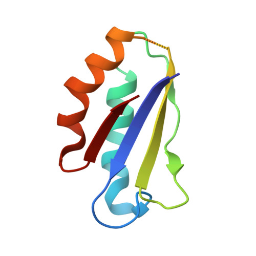
    4Y0L

  • PubMed Abstract: 

    Mycobacterium tuberculosis mycobacterial membrane protein large (MmpL) proteins are important in substrate transport across the inner membrane. Here, we show that MmpL proteins are classified into two phylogenetic clusters, where MmpL cluster II contains three soluble domains (D1, D2, and D3) and has two full-length members, MmpL3 and MmpL11. Significantly, MmpL3 is currently the most druggable M. tuberculosis target. We have solved the 2.4-Å MmpL11-D2 crystal structure, revealing structural homology to periplasmic porter subdomains of RND (multidrug) transporters. The resulting predicted cluster II MmpL membrane topology has D1 and D2 residing, and possibly interacting, within the periplasm. Crosslinking and biolayer interferometry experiments confirm that cluster II D1 and D2 bind with weak affinities, and guided D1-D2 heterodimeric model assemblies. The predicted full-length MmpL3 and MmpL11 structural models reveal key substrate binding and transport residues, and may serve as templates to set the stage for in silico anti-tuberculosis drug development.


  • Organizational Affiliation
    • Department of Molecular Biology and Biochemistry, UCI, Irvine, CA 92697, USA.

Macromolecule Content 

  • Total Structure Weight: 10.41 kDa 
  • Atom Count: 612 
  • Modeled Residue Count: 80 
  • Deposited Residue Count: 88 
  • Unique protein chains: 1

Macromolecules

Find similar proteins by:|  3D Structure
Entity ID: 1
MoleculeChains  Sequence LengthOrganismDetailsImage
Putative membrane protein mmpL1188Mycobacterium tuberculosis H37RvMutation(s): 0 
Gene Names: mmpL11Rv0202cMTV033.10c
UniProt
Find proteins for P9WJT9 (Mycobacterium tuberculosis (strain ATCC 25618 / H37Rv))
Explore P9WJT9 
Go to UniProtKB:  P9WJT9
Entity Groups
Sequence Clusters30% Identity50% Identity70% Identity90% Identity95% Identity100% Identity
UniProt GroupP9WJT9
Sequence Annotations
Expand
Reference Sequence

Small Molecules

Ligands 2 Unique
IDChains Name / Formula / InChI Key2D Diagram3D Interactions
IOD

Query on IOD


Download:Ideal Coordinates CCD File
B [auth A]
C [auth A]
D [auth A]
E [auth A]
F [auth A]
B [auth A],
C [auth A],
D [auth A],
E [auth A],
F [auth A],
G [auth A],
H [auth A],
I [auth A],
J [auth A]
IODIDE ION
I
XMBWDFGMSWQBCA-UHFFFAOYSA-M
SO4

Query on SO4


Download:Ideal Coordinates CCD File
K [auth A]SULFATE ION
O4 S
QAOWNCQODCNURD-UHFFFAOYSA-L

Experimental Data & Validation

Experimental Data

  • Method: X-RAY DIFFRACTION
  • Resolution: 2.40 Å
  • R-Value Free:  0.254 (Depositor), 0.258 (DCC) 
  • R-Value Work:  0.208 (Depositor), 0.213 (DCC) 
  • R-Value Observed: 0.212 (Depositor) 
Space Group: C 2 2 21
Unit Cell:
Length ( Å )Angle ( ˚ )
a = 65.6α = 90
b = 91.73β = 90
c = 32.85γ = 90
Software Package:
Software NamePurpose
iMOSFLMdata reduction
PHENIXphasing
PHENIXrefinement
Cootmodel building
PDB_EXTRACTdata extraction

Structure Validation

View Full Validation Report


Entry History 

Deposition Data

Revision History  (Full details and data files)

  • Version 1.0: 2015-08-12
    Type: Initial release
  • Version 1.1: 2015-09-02
    Changes: Database references, Other
  • Version 1.2: 2024-02-28
    Changes: Data collection, Database references, Derived calculations