4XYF | pdb_00004xyf

Crystal structure of c-Met in complex with (S)-5-(8-fluoro-3-(1-(3-(2-methoxyethoxy)quinolin-6-yl)ethyl)-[1,2,4]triazolo[4,3-a]pyridin-6-yl)-3-methylisoxazole


Experimental Data Snapshot

  • Method: X-RAY DIFFRACTION
  • Resolution: 1.85 Å
  • R-Value Free: 
    0.236 (Depositor), 0.232 (DCC) 
  • R-Value Work: 
    0.186 (Depositor), 0.186 (DCC) 
  • R-Value Observed: 
    0.188 (Depositor) 

Starting Model: experimental
View more details

wwPDB Validation 3D Report Full Report

Validation slider image for 4XYF

Ligand Structure Quality Assessment 


This is version 1.2 of the entry. See complete history

Literature

Discovery of Potent and Selective 8-Fluorotriazolopyridine c-Met Inhibitors.

Peterson, E.A.Teffera, Y.Albrecht, B.K.Bauer, D.Bellon, S.F.Boezio, A.Boezio, C.Broome, M.A.Choquette, D.Copeland, K.W.Dussault, I.Lewis, R.Lin, M.H.Lohman, J.Liu, J.Potashman, M.Rex, K.Shimanovich, R.Whittington, D.A.Vaida, K.R.Harmange, J.C.

(2015) J Med Chem 58: 2417-2430

  • DOI: https://doi.org/10.1021/jm501913a
  • Primary Citation Related Structures: 
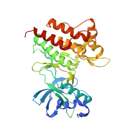
    4XMO, 4XYF

  • PubMed Abstract: 

    The overexpression of c-Met and/or hepatocyte growth factor (HGF), the amplification of the MET gene, and mutations in the c-Met kinase domain can activate signaling pathways that contribute to cancer progression by enabling tumor cell proliferation, survival, invasion, and metastasis. Herein, we report the discovery of 8-fluorotriazolopyridines as inhibitors of c-Met activity. Optimization of the 8-fluorotriazolopyridine scaffold through the combination of structure-based drug design, SAR studies, and metabolite identification provided potent (cellular IC50 < 10 nM), selective inhibitors of c-Met with desirable pharmacokinetic properties that demonstrate potent inhibition of HGF-mediated c-Met phosphorylation in a mouse liver pharmacodynamic model.


  • Organizational Affiliation
    • Amgen Inc. , 360 Binney Street, Cambridge, Massachusetts 02142, United States.

Macromolecule Content 

  • Total Structure Weight: 35.66 kDa 
  • Atom Count: 2,613 
  • Modeled Residue Count: 290 
  • Deposited Residue Count: 309 
  • Unique protein chains: 1

Macromolecules

Find similar proteins by:|  3D Structure
Entity ID: 1
MoleculeChains  Sequence LengthOrganismDetailsImage
Hepatocyte growth factor receptor309Homo sapiensMutation(s): 0 
Gene Names: MET
EC: 2.7.10.1
UniProt & NIH Common Fund Data Resources
Find proteins for P08581 (Homo sapiens)
Explore P08581 
Go to UniProtKB:  P08581
PHAROS:  P08581
GTEx:  ENSG00000105976 
Entity Groups
Sequence Clusters30% Identity50% Identity70% Identity90% Identity95% Identity100% Identity
UniProt GroupP08581
Sequence Annotations
Expand
Reference Sequence

Small Molecules

Ligands 1 Unique
IDChains Name / Formula / InChI Key2D Diagram3D Interactions
44X

Query on 44X


Download:Ideal Coordinates CCD File
B [auth A]6-{(1S)-1-[8-fluoro-6-(3-methyl-1,2-oxazol-5-yl)[1,2,4]triazolo[4,3-a]pyridin-3-yl]ethyl}-3-(2-methoxyethoxy)quinoline
C24 H22 F N5 O3
SCRRRLBPWUHHER-HNNXBMFYSA-N

Experimental Data & Validation

Experimental Data

  • Method: X-RAY DIFFRACTION
  • Resolution: 1.85 Å
  • R-Value Free:  0.236 (Depositor), 0.232 (DCC) 
  • R-Value Work:  0.186 (Depositor), 0.186 (DCC) 
  • R-Value Observed: 0.188 (Depositor) 
Space Group: P 21 21 21
Unit Cell:
Length ( Å )Angle ( ˚ )
a = 41.893α = 90
b = 43.406β = 90
c = 157.978γ = 90
Software Package:
Software NamePurpose
CrystalCleardata collection
DENZOdata reduction
SCALEPACKdata scaling
PDB_EXTRACTdata extraction
EPMRphasing
REFMACrefinement

Structure Validation

View Full Validation Report


Ligand Structure Quality Assessment 


Entry History 

Deposition Data

Revision History  (Full details and data files)

  • Version 1.0: 2015-03-11
    Type: Initial release
  • Version 1.1: 2015-03-25
    Changes: Database references
  • Version 1.2: 2023-09-27
    Changes: Data collection, Database references, Derived calculations, Refinement description, Source and taxonomy, Structure summary