Nanomolar inhibitors of Mycobacterium tuberculosis glutamine synthetase 1: Synthesis, biological evaluation and X-ray crystallographic studies.
Couturier, C., Silve, S., Morales, R., Pessegue, B., Llopart, S., Nair, A., Bauer, A., Scheiper, B., Poverlein, C., Ganzhorn, A., Lagrange, S., Bacque, E.(2015) Bioorg Med Chem Lett 25: 1455-1459
- PubMed: 25770781 Search on PubMed
- DOI: https://doi.org/10.1016/j.bmcl.2015.02.035
- Primary Citation Related Structures:
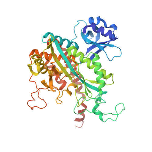
4XYC - PubMed Abstract:
A series of imidazo[1,2-a]indeno[1,2-e]pyrazin-4-ones that potently inhibit M. tuberculosis glutamine synthetase (GlnA1) has been identified by high throughput screening. Exploration of this series was performed owing to a short chemistry program. Despite possibly nanomolar inhibitions, none of these compounds was active on whole cell Mtb, suggesting that GlnA1 may not be a suitable target to find new anti-tubercular drugs.
- Sanofi R&D, TSU Infectious Disease, 195, route d'Espagne BP 13669, 31036 Toulouse Cedex 1, France. Electronic address: cedric.couturier@sanofi.com.
Organizational Affiliation: