4XY3

Structure of ESX-1 secreted protein EspB


Experimental Data Snapshot

  • Method: X-RAY DIFFRACTION
  • Resolution: 3.04 Å
  • R-Value Free: 0.266 
  • R-Value Work: 0.220 
  • R-Value Observed: 0.222 

wwPDB Validation   3D Report Full Report


This is version 1.6 of the entry. See complete history


Literature

Structure of EspB, a secreted substrate of the ESX-1 secretion system of Mycobacterium tuberculosis.

Korotkova, N.Piton, J.Wagner, J.M.Boy-Rottger, S.Japaridze, A.Evans, T.J.Cole, S.T.Pojer, F.Korotkov, K.V.

(2015) J Struct Biol 191: 236-244

  • DOI: https://doi.org/10.1016/j.jsb.2015.06.003
  • Primary Citation of Related Structures:  
    4XWP, 4XXN, 4XXX, 4XY3

  • PubMed Abstract: 

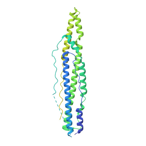
    Mycobacterium tuberculosis secretes multiple virulence factors during infection via the general Sec and Tat pathways, and via specialized ESX secretion systems, also referred to as type VII secretion systems. The ESX-1 secretion system is an important virulence determinant because deletion of ESX-1 leads to attenuation of M. tuberculosis. ESX-1 secreted protein B (EspB) contains putative PE (Pro-Glu) and PPE (Pro-Pro-Glu) domains, and a C-terminal domain, which is processed by MycP1 protease during secretion. We determined the crystal structure of PE-PPE domains of EspB, which represents an all-helical, elongated molecule closely resembling the structure of the PE25-PPE41 heterodimer despite limited sequence similarity. Also, we determined the structure of full-length EspB, which does not have interpretable electron density for the C-terminal domain confirming that it is largely disordered. Comparative analysis of EspB in cell lysate and culture filtrates of M. tuberculosis revealed that mature secreted EspB forms oligomers. Electron microscopy analysis showed that the N-terminal fragment of EspB forms donut-shaped particles. These data provide a rationale for the future investigation of EspB's role in M. tuberculosis pathogenesis.


  • Organizational Affiliation

    Department of Molecular & Cellular Biochemistry, and Center for Structural Biology, University of Kentucky, Lexington, KY, 40536, United States.


Macromolecules
Find similar proteins by:  (by identity cutoff)  |  3D Structure
Entity ID: 1
MoleculeChains Sequence LengthOrganismDetailsImage
ESX-1 secretion-associated protein EspB462Mycobacterium tuberculosis H37RvMutation(s): 0 
Gene Names: espBmtb48Rv3881cMTV027.16c
UniProt
Find proteins for P9WJD9 (Mycobacterium tuberculosis (strain ATCC 25618 / H37Rv))
Explore P9WJD9 
Go to UniProtKB:  P9WJD9
Entity Groups  
Sequence Clusters30% Identity50% Identity70% Identity90% Identity95% Identity100% Identity
UniProt GroupP9WJD9
Sequence Annotations
Expand
  • Reference Sequence
Experimental Data & Validation

Experimental Data

  • Method: X-RAY DIFFRACTION
  • Resolution: 3.04 Å
  • R-Value Free: 0.266 
  • R-Value Work: 0.220 
  • R-Value Observed: 0.222 
  • Space Group: C 2 2 21
Unit Cell:
Length ( Å )Angle ( ˚ )
a = 72.11α = 90
b = 146.49β = 90
c = 94.22γ = 90
Software Package:
Software NamePurpose
XDSdata reduction
PHASERphasing
REFMACrefinement
PDB_EXTRACTdata extraction
XSCALEdata scaling

Structure Validation

View Full Validation Report


Entry History & Funding Information

Deposition Data


Funding OrganizationLocationGrant Number
National Institutes of Health/National Institute of General Medical Sciences (NIH/NIGMS)United StatesP20GM103486

Revision History  (Full details and data files)

  • Version 1.0: 2015-02-18
    Type: Initial release
  • Version 1.1: 2015-02-25
    Changes: Structure summary
  • Version 1.2: 2015-06-17
    Changes: Database references
  • Version 1.3: 2015-08-05
    Changes: Database references
  • Version 1.4: 2017-09-06
    Changes: Author supporting evidence, Database references, Derived calculations
  • Version 1.5: 2019-12-25
    Changes: Author supporting evidence
  • Version 1.6: 2023-09-27
    Changes: Data collection, Database references, Refinement description