4XXR | pdb_00004xxr

Atomic Resolution X-Ray Crystal Structure of a Ruthenocene Conjugated Beta-Lactam Antibiotic in Complex with CTX-M-14 E166A Beta-Lactamase


Experimental Data Snapshot

  • Method: X-RAY DIFFRACTION
  • Resolution: 1.18 Å
  • R-Value Free: 
    0.153 (Depositor), 0.161 (DCC) 
  • R-Value Work: 
    0.125 (Depositor), 0.135 (DCC) 
  • R-Value Observed: 
    0.126 (Depositor) 

Starting Model: experimental
View more details

wwPDB Validation 3D Report Full Report

Validation slider image for 4XXR

Ligand Structure Quality Assessment 


This is version 1.6 of the entry. See complete history

Literature

Antibacterial properties and atomic resolution X-ray complex crystal structure of a ruthenocene conjugated beta-lactam antibiotic.

Lewandowski, E.M.Skiba, J.Torelli, N.J.Rajnisz, A.Solecka, J.Kowalski, K.Chen, Y.

(2015) Chem Commun (Camb) 51: 6186-6189

  • DOI: https://doi.org/10.1039/c5cc00904a
  • Primary Citation Related Structures: 
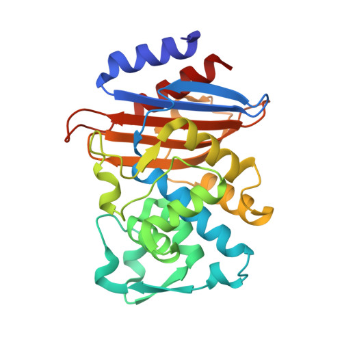
    4XXR

  • PubMed Abstract: 

    We have determined a 1.18 Å resolution X-ray crystal structure of a novel ruthenocenyle-6-aminopenicillinic acid in complex with CTX-M β-lactamase, showing unprecedented details of interactions between ruthenocene and protein. As the first product complex with an intact catalytic serine, the structure also offers insights into β-lactamase catalysis and inhibitor design.


  • Organizational Affiliation
    • Department of Molecular Medicine, University of South Florida, Tampa, Florida 33612, USA. ychen1@health.usf.edu.

Macromolecule Content 

  • Total Structure Weight: 59.11 kDa 
  • Atom Count: 5,221 
  • Modeled Residue Count: 522 
  • Deposited Residue Count: 526 
  • Unique protein chains: 1

Macromolecules

Find similar proteins by:|  3D Structure
Entity ID: 1
MoleculeChains  Sequence LengthOrganismDetailsImage
CTX-M-14 Class A Beta-Lactamase
A, B
263Escherichia coliMutation(s): 1 
Gene Names: 
EC: 3.5.2.6
UniProt
Find proteins for Q9L5C7 (Escherichia coli)
Explore Q9L5C7 
Go to UniProtKB:  Q9L5C7
Entity Groups
Sequence Clusters30% Identity50% Identity70% Identity90% Identity95% Identity100% Identity
UniProt GroupQ9L5C7
Sequence Annotations
Expand
Reference Sequence

Small Molecules

Ligands 4 Unique
IDChains Name / Formula / InChI Key2D Diagram3D Interactions
JSE

Query on JSE


Download:Ideal Coordinates CCD File
F [auth A][(1,2,3,4,5-eta)-1-(4-{[carboxy(4-carboxy-5,5-dimethyl-1,3-thiazolidin-2-yl)methyl]amino}-4-oxobutanoyl)cyclopentadienyl][(1,2,3,4,5-eta)-cyclopentadienyl]ruthenium
C22 H17 N2 O6 Ru S
DONWCTWXNJLLEK-PAALKQLMSA-N
JSC

Query on JSC


Download:Ideal Coordinates CCD File
E [auth A],
G [auth A],
J [auth B]
[(1,2,3,4,5-eta)-1-(4-{[carboxy(4-carboxy-5,5-dimethyl-1,3-thiazolidin-2-yl)methyl]amino}-4-oxobutanoyl)cyclopentadienyl][(1,2,3,4,5-eta)-cyclopentadienyl]ruthenium
C22 H17 N2 O6 Ru S
DONWCTWXNJLLEK-HTTVLNLHSA-N
JSD

Query on JSD


Download:Ideal Coordinates CCD File
D [auth A],
I [auth B]
[(1,2,3,4,5-eta)-1-(4-{[(4-carboxy-5,5-dimethyl-1,3-thiazolidin-2-yl)methyl]amino}-4-oxobutanoyl)cyclopentadienyl][(1,2,3,4,5-eta)-cyclopentadienyl]ruthenium
C21 H17 N2 O4 Ru S
JRWKHIBMJXULTB-BQFBZIMZSA-N
K

Query on K


Download:Ideal Coordinates CCD File
C [auth A],
H [auth B]
POTASSIUM ION
K
NPYPAHLBTDXSSS-UHFFFAOYSA-N

Experimental Data & Validation

Experimental Data

  • Method: X-RAY DIFFRACTION
  • Resolution: 1.18 Å
  • R-Value Free:  0.153 (Depositor), 0.161 (DCC) 
  • R-Value Work:  0.125 (Depositor), 0.135 (DCC) 
  • R-Value Observed: 0.126 (Depositor) 
Space Group: P 1 21 1
Unit Cell:
Length ( Å )Angle ( ˚ )
a = 44.899α = 90
b = 106.775β = 101.87
c = 47.903γ = 90
Software Package:
Software NamePurpose
REFMACrefinement
HKL-2000data scaling
SCALEPACKdata scaling
DENZOdata reduction
PHASERphasing

Structure Validation

View Full Validation Report


Ligand Structure Quality Assessment 


Entry History 

& Funding Information

Deposition Data


Funding OrganizationLocationGrant Number
National Institutes of Health/National Institute Of Allergy and Infectious Diseases (NIH/NIAID)United StatesAI 103158

Revision History  (Full details and data files)

  • Version 1.0: 2015-03-18
    Type: Initial release
  • Version 1.1: 2015-04-08
    Changes: Database references
  • Version 1.2: 2016-03-09
    Changes: Experimental preparation
  • Version 1.3: 2017-09-06
    Changes: Author supporting evidence, Derived calculations
  • Version 1.4: 2017-11-22
    Changes: Refinement description
  • Version 1.5: 2019-12-11
    Changes: Author supporting evidence
  • Version 1.6: 2023-09-27
    Changes: Data collection, Database references, Derived calculations, Refinement description