Small-Molecule Allosteric Modulators of the Protein Kinase PDK1 from Structure-Based Docking.
Rettenmaier, T.J., Fan, H., Karpiak, J., Doak, A., Sali, A., Shoichet, B.K., Wells, J.A.(2015) J Med Chem 58: 8285-8291
- PubMed: 26443011 Search on PubMedSearch on PubMed Central
- DOI: https://doi.org/10.1021/acs.jmedchem.5b01216
- Primary Citation Related Structures:
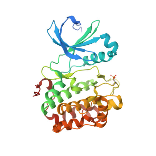
4XX9 - PubMed Abstract:
Finding small molecules that target allosteric sites remains a grand challenge for ligand discovery. In the protein kinase field, only a handful of highly selective allosteric modulators have been found. Thus, more general methods are needed to discover allosteric modulators for additional kinases. Here, we use virtual screening against an ensemble of both crystal structures and comparative models to identify ligands for an allosteric peptide-binding site on the protein kinase PDK1 (the PIF pocket). We optimized these ligands through an analog-by-catalog search that yielded compound 4, which binds to PDK1 with 8 μM affinity. We confirmed the docking poses by determining a crystal structure of PDK1 in complex with 4. Because the PIF pocket appears to be a recurring structural feature of the kinase fold, known generally as the helix αC patch, this approach may enable the discovery of allosteric modulators for other kinases.
- Chemistry and Chemical Biology Graduate Program, University of California, San Francisco, California 94158, United States.
Organizational Affiliation: