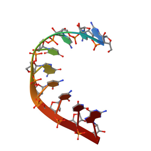
Watson-Crick-like pairs in CCUG repeats: evidence for tautomeric shifts or protonation.
Rypniewski, W., Banaszak, K., Kulinski, T., Kiliszek, A.(2016) RNA 22: 22-31
- PubMed: 26543073 Search on PubMedSearch on PubMed Central
- DOI: https://doi.org/10.1261/rna.052399.115
- Primary Citation Related Structures:
4XW0, 4XW1 - PubMed Abstract:
RNA transcripts that include expanded CCUG repeats are associated with myotonic dystrophy type 2. Crystal structures of two CCUG-containing oligomers show that the RNA strands associate into slipped duplexes that contain noncanonical C-U pairs that have apparently undergone tautomeric transition or protonation resulting in an unusual Watson-Crick-like pairing. The overhanging ends of the duplexes interact forming U-U pairs, which also show tautomerism. Duplexes consisting of CCUG repeats are thermodynamically less stable than the trinucleotide repeats involved in the TRED genetic disorders, but introducing LNA residues increases their stability and raises the melting temperature of the studied oligomers by ∼10°C, allowing detailed crystallographic studies. Quantum mechanical calculations were performed to test the possibility of the tautomeric transitions or protonation within the noncanonical pairs. The results indicate that tautomeric or ionic shifts of nucleobases can manifest themselves in biological systems, supplementing the canonical "rules of engagement."
- Institute of Bioorganic Chemistry, Polish Academy of Sciences, 61-704 Poznan, Poland.
Organizational Affiliation: