4XU0

Mycobacterium tuberculosis biotin ligase complexed with bisubstrate inhibitor 73 that has a 2'-methyl on the ribose


Experimental Data Snapshot

  • Method: X-RAY DIFFRACTION
  • Resolution: 1.60 Å
  • R-Value Free: 0.220 
  • R-Value Work: 0.195 
  • R-Value Observed: 0.196 

wwPDB Validation   3D Report Full Report


Ligand Structure Quality Assessment 


This is version 1.4 of the entry. See complete history


Literature

Targeting Mycobacterium tuberculosis Biotin Protein Ligase (MtBPL) with Nucleoside-Based Bisubstrate Adenylation Inhibitors.

Bockman, M.R.Kalinda, A.S.Petrelli, R.De la Mora-Rey, T.Tiwari, D.Liu, F.Dawadi, S.Nandakumar, M.Rhee, K.Y.Schnappinger, D.Finzel, B.C.Aldrich, C.C.

(2015) J Med Chem 58: 7349-7369

  • DOI: https://doi.org/10.1021/acs.jmedchem.5b00719
  • Primary Citation of Related Structures:  
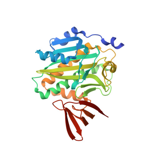
    4XTV, 4XTW, 4XTX, 4XTY, 4XTZ, 4XU0, 4XU1, 4XU2, 4XU3

  • PubMed Abstract: 

    Mycobacterium tuberculosis (Mtb), responsible for both latent and symptomatic tuberculosis (TB), remains the second leading cause of mortality among infectious diseases worldwide. Mycobacterial biotin protein ligase (MtBPL) is an essential enzyme in Mtb and regulates lipid metabolism through the post-translational biotinylation of acyl coenzyme A carboxylases. We report the synthesis and evaluation of a systematic series of potent nucleoside-based inhibitors of MtBPL that contain modifications to the ribofuranosyl ring of the nucleoside. All compounds were characterized by isothermal titration calorimetry (ITC) and shown to bind potently with K(D)s ≤ 2 nM. Additionally, we obtained high-resolution cocrystal structures for a majority of the compounds. Despite fairly uniform biochemical potency, the whole-cell Mtb activity varied greatly with minimum inhibitory concentrations (MIC) ranging from 0.78 to >100 μM. Cellular accumulation studies showed a nearly 10-fold enhancement in accumulation of a C-2'-α analogue over the corresponding C-2'-β analogue, consistent with their differential whole-cell activity.


  • Organizational Affiliation

    Department of Medicinal Chemistry, University of Minnesota, Minneapolis, MN 55455, USA.


Macromolecules
Find similar proteins by:  (by identity cutoff)  |  3D Structure
Entity ID: 1
MoleculeChains Sequence LengthOrganismDetailsImage
Bifunctional ligase/repressor BirA
A, B
270Mycobacterium tuberculosisMutation(s): 0 
Gene Names: 
EC: 6.3.4.15
UniProt
Find proteins for I6YFP0 (Mycobacterium tuberculosis (strain ATCC 25618 / H37Rv))
Explore I6YFP0 
Go to UniProtKB:  I6YFP0
Entity Groups  
Sequence Clusters30% Identity50% Identity70% Identity90% Identity95% Identity100% Identity
UniProt GroupI6YFP0
Sequence Annotations
Expand
  • Reference Sequence
Small Molecules
Ligands 1 Unique
IDChains Name / Formula / InChI Key2D Diagram3D Interactions
44N
Query on 44N

Download Ideal Coordinates CCD File 
C [auth A],
D [auth B]
2'-C-methyl-5'-O-({5-[(3aS,4S,6aR)-2-oxohexahydro-1H-thieno[3,4-d]imidazol-4-yl]pentanoyl}sulfamoyl)adenosine
C21 H30 N8 O8 S2
BGUXFAITPCEERX-BKJQVYKZSA-N
Binding Affinity Annotations 
IDSourceBinding Affinity
44N Binding MOAD:  4XU0 Kd: 0.77 (nM) from 1 assay(s)
Experimental Data & Validation

Experimental Data

  • Method: X-RAY DIFFRACTION
  • Resolution: 1.60 Å
  • R-Value Free: 0.220 
  • R-Value Work: 0.195 
  • R-Value Observed: 0.196 
  • Space Group: P 21 21 21
Unit Cell:
Length ( Å )Angle ( ˚ )
a = 63.55α = 90
b = 68.907β = 90
c = 115.333γ = 90
Software Package:
Software NamePurpose
PHASERphasing
PHENIXrefinement
PDB_EXTRACTdata extraction
SCALAdata scaling

Structure Validation

View Full Validation Report


Ligand Structure Quality Assessment 


Entry History & Funding Information

Deposition Data


Funding OrganizationLocationGrant Number
National Institutes of Health/National Institute Of Allergy and Infectious Diseases (NIH/NIAID)United StatesR01 AI091790-01

Revision History  (Full details and data files)

  • Version 1.0: 2015-09-02
    Type: Initial release
  • Version 1.1: 2015-10-07
    Changes: Database references, Experimental preparation
  • Version 1.2: 2017-09-06
    Changes: Author supporting evidence, Derived calculations
  • Version 1.3: 2019-12-11
    Changes: Author supporting evidence
  • Version 1.4: 2023-09-27
    Changes: Data collection, Database references, Refinement description