4XSG | pdb_00004xsg

The complex structure of C3cer exoenzyme and GTP bound RhoA (NADH-free state)


Experimental Data Snapshot

  • Method: X-RAY DIFFRACTION
  • Resolution: 1.80 Å
  • R-Value Free: 
    0.223 (Depositor), 0.231 (DCC) 
  • R-Value Work: 
    0.175 (Depositor), 0.182 (DCC) 
  • R-Value Observed: 
    0.177 (Depositor) 

Starting Models: experimental
View more details

wwPDB Validation 3D Report Full Report

Validation slider image for 4XSG

Ligand Structure Quality Assessment 


This is version 1.3 of the entry. See complete history

Literature

Rho GTPase Recognition by C3 Exoenzyme Based on C3-RhoA Complex Structure.

Toda, A.Tsurumura, T.Yoshida, T.Tsumori, Y.Tsuge, H.

(2015) J Biological Chem 290: 19423-19432

  • DOI: https://doi.org/10.1074/jbc.M115.653220
  • Primary Citation Related Structures: 
    4XSG, 4XSH, 5BWM

  • PubMed Abstract: 

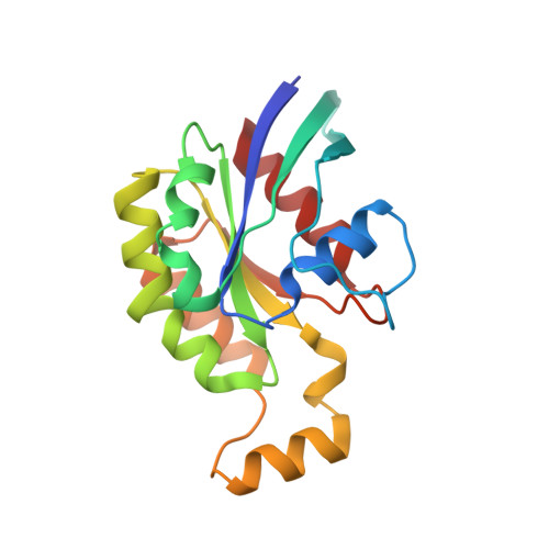
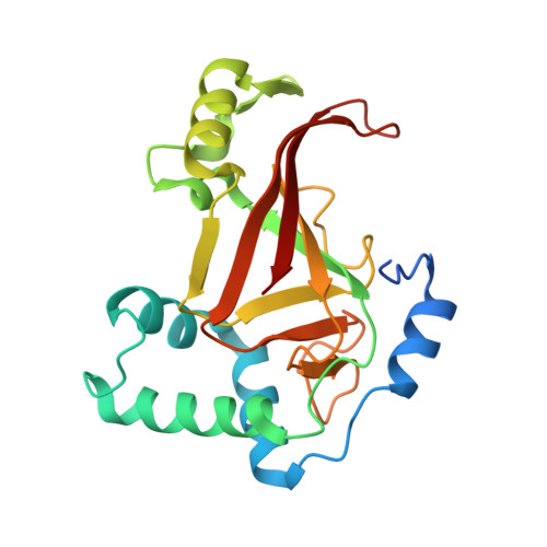
    C3 exoenzyme is a mono-ADP-ribosyltransferase (ART) that catalyzes transfer of an ADP-ribose moiety from NAD(+) to Rho GTPases. C3 has long been used to study the diverse regulatory functions of Rho GTPases. How C3 recognizes its substrate and how ADP-ribosylation proceeds are still poorly understood. Crystal structures of C3-RhoA complex reveal that C3 recognizes RhoA via the switch I, switch II, and interswitch regions. In C3-RhoA(GTP) and C3-RhoA(GDP), switch I and II adopt the GDP and GTP conformations, respectively, which explains why C3 can ADP-ribosylate both nucleotide forms. Based on structural information, we successfully changed Cdc42 to an active substrate with combined mutations in the C3-Rho GTPase interface. Moreover, the structure reflects the close relationship among Gln-183 in the QXE motif (C3), a modified Asn-41 residue (RhoA) and NC1 of NAD(H), which suggests that C3 is the prototype ART. These structures show directly for the first time that the ARTT loop is the key to target protein recognition, and they also serve to bridge the gaps among independent studies of Rho GTPases and C3.


  • Organizational Affiliation
    • From the Department of Bioresource and Environmental Sciences, Faculty of Life Sciences, and.

Macromolecule Content 

  • Total Structure Weight: 46.21 kDa 
  • Atom Count: 3,418 
  • Modeled Residue Count: 381 
  • Deposited Residue Count: 398 
  • Unique protein chains: 2

Macromolecules

Find similar proteins by:|  3D Structure
Entity ID: 1
MoleculeChains  Sequence LengthOrganismDetailsImage
Transforming protein RhoA179Homo sapiensMutation(s): 1 
Gene Names: RHOAARH12ARHARHO12
EC: 3.6.5.2
UniProt & NIH Common Fund Data Resources
Find proteins for P61586 (Homo sapiens)
Explore P61586 
Go to UniProtKB:  P61586
PHAROS:  P61586
GTEx:  ENSG00000067560 
Entity Groups
Sequence Clusters30% Identity50% Identity70% Identity90% Identity95% Identity100% Identity
UniProt GroupP61586
Sequence Annotations
Expand
Reference Sequence
Find similar proteins by:|  3D Structure
Entity ID: 2
MoleculeChains  Sequence LengthOrganismDetailsImage
ADP-ribosyltransferase219Bacillus cereusMutation(s): 0 
Gene Names: c3cer
UniProt
Find proteins for Q8KNY0 (Bacillus cereus)
Explore Q8KNY0 
Go to UniProtKB:  Q8KNY0
Entity Groups
Sequence Clusters30% Identity50% Identity70% Identity90% Identity95% Identity100% Identity
UniProt GroupQ8KNY0
Sequence Annotations
Expand
Reference Sequence

Experimental Data & Validation

Experimental Data

  • Method: X-RAY DIFFRACTION
  • Resolution: 1.80 Å
  • R-Value Free:  0.223 (Depositor), 0.231 (DCC) 
  • R-Value Work:  0.175 (Depositor), 0.182 (DCC) 
  • R-Value Observed: 0.177 (Depositor) 
Space Group: P 32
Unit Cell:
Length ( Å )Angle ( ˚ )
a = 50.745α = 90
b = 50.745β = 90
c = 135.871γ = 120
Software Package:
Software NamePurpose
REFMACrefinement
HKL-2000data reduction
HKL-2000data scaling
PHENIXphasing

Structure Validation

View Full Validation Report


Ligand Structure Quality Assessment 


Entry History 

& Funding Information

Deposition Data


Funding OrganizationLocationGrant Number
Grants-in-Aid for Scientific Research, MEXT of JapanJapan25121733

Revision History  (Full details and data files)

  • Version 1.0: 2015-06-24
    Type: Initial release
  • Version 1.1: 2015-08-19
    Changes: Database references
  • Version 1.2: 2020-02-05
    Changes: Data collection, Database references, Derived calculations
  • Version 1.3: 2023-11-08
    Changes: Data collection, Database references, Refinement description