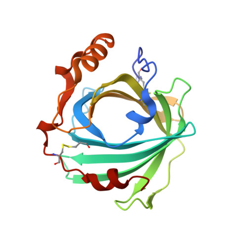
Structure and dynamics of the membrane attaching nitric oxide transporter nitrophorin 7.
Knipp, M., Ogata, H., Soavi, G., Cerullo, G., Allegri, A., Abbruzzetti, S., Bruno, S., Viappiani, C., Bidon-Chanal, A., Luque, F.J.(2015) F1000Res 4: 45-45
- PubMed: 26167269 Search on PubMedSearch on PubMed Central
- DOI: https://doi.org/10.12688/f1000research.6060.1
- Primary Citation Related Structures:
4XMC, 4XMD, 4XME, 4XMF, 4XMG, 4XMH - PubMed Abstract:
Nitrophorins represent a unique class of heme proteins that are able to perform the delicate transportation and release of the free-radical gaseous messenger nitric oxide (NO) in a pH-triggered manner. Besides its ability to bind to phospholipid membranes, the N-terminus contains an additional Leu-Pro-Gly stretch, which is a unique sequence trait, and the heme cavity is significantly altered with respect to other nitrophorins. These distinctive features encouraged us to solve the X-ray crystallographic structures of NP7 at low and high pH and bound with different heme ligands (nitric oxide, histamine, imidazole). The overall fold of the lipocalin motif is well preserved in the different X-ray structures and resembles the fold of other nitrophorins. However, a chain-like arrangement in the crystal lattice due to a number of head-to-tail electrostatic stabilizing interactions is found in NP7. Furthermore, the X-ray structures also reveal ligand-dependent changes in the orientation of the heme, as well as in specific interactions between the A-B and G-H loops, which are considered to be relevant for the biological function of nitrophorins. Fast and ultrafast laser triggered ligand rebinding experiments demonstrate the pH-dependent ligand migration within the cavities and the exit route. Finally, the topological distribution of pockets located around the heme as well as from inner cavities present at the rear of the protein provides a distinctive feature in NP7, so that while a loop gated exit mechanism to the solvent has been proposed for most nitrophorins, a more complex mechanism that involves several interconnected gas hosting cavities is proposed for NP7.
- Max-Planck-Institut für Chemische Energiekonversion, Mülheim an der Ruhr, 45470, Germany.
Organizational Affiliation: