


Crystal structure of human NLRP12 PYD domain and implication in homotypic interaction
Jin, T., Huang, M., Jiang, J., Smith, P., Xiao, T.To be published.
Experimental Data Snapshot
Starting Models: experimental
View more details
wwPDB Validation 3D Report Full Report

Entity ID: 1 | |||||
|---|---|---|---|---|---|
| Molecule | Chains | Sequence Length | Organism | Details | Image |
| Maltose-binding periplasmic protein,NACHT, LRR and PYD domains-containing protein 12 | 480 | Escherichia coli O157:H7, Homo sapiens This entity is chimeric | Mutation(s): 0 Gene Names: malE, Z5632, ECs5017, NLRP12, NALP12, PYPAF7, RNO | 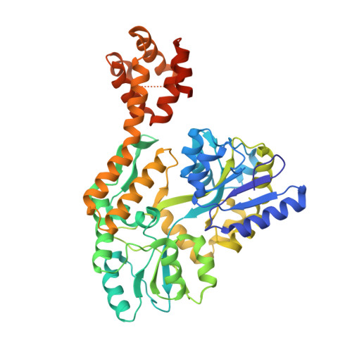 | |
UniProt & NIH Common Fund Data Resources | |||||
PHAROS: P59046 GTEx: ENSG00000142405 | |||||
Entity Groups | |||||
| Sequence Clusters | 30% Identity50% Identity70% Identity90% Identity95% Identity100% Identity | ||||
| UniProt Group | P59046 | ||||
Sequence AnnotationsExpand | |||||
Reference Sequence | |||||
| Ligands 2 Unique | |||||
|---|---|---|---|---|---|
| ID | Chains | Name / Formula / InChI Key | 2D Diagram | 3D Interactions | |
| FMT Download:Ideal Coordinates CCD File | E [auth A], F [auth A], I [auth B], J [auth B] | FORMIC ACID C H2 O2 BDAGIHXWWSANSR-UHFFFAOYSA-N |  | ||
| NA Download:Ideal Coordinates CCD File | G [auth A] H [auth A] K [auth B] L [auth B] M [auth B] | SODIUM ION Na FKNQFGJONOIPTF-UHFFFAOYSA-N |  | ||
Entity ID: 2 | |||||
|---|---|---|---|---|---|
| ID | Chains | Name | Type/Class | 2D Diagram | 3D Interactions |
| PRD_900001 Query on PRD_900001 | C, D | alpha-maltose | Oligosaccharide / Nutrient |  |
| Length ( Å ) | Angle ( ˚ ) |
|---|---|
| a = 52.72 | α = 90 |
| b = 103.62 | β = 90 |
| c = 186.74 | γ = 90 |
| Software Name | Purpose |
|---|---|
| PHENIX | refinement |
| HKL-2000 | data reduction |
| HKL-2000 | data scaling |
| PHASER | phasing |


