4XCZ

X-ray structure of the N-formyltransferase QdtF from Providencia alcalifaciens in complex with TDP-Qui3n and N5-THF


Experimental Data Snapshot

  • Method: X-RAY DIFFRACTION
  • Resolution: 1.50 Å
  • R-Value Free: 0.215 
  • R-Value Work: 0.183 
  • R-Value Observed: 0.185 

wwPDB Validation   3D Report Full Report


Ligand Structure Quality Assessment 


This is version 1.3 of the entry. See complete history


Literature

New Role for the Ankyrin Repeat Revealed by a Study of the N-Formyltransferase from Providencia alcalifaciens.

Woodford, C.R.Thoden, J.B.Holden, H.M.

(2015) Biochemistry 54: 631-638

  • DOI: https://doi.org/10.1021/bi501539a
  • Primary Citation of Related Structures:  
    4XCZ, 4XD0, 4XD1

  • PubMed Abstract: 

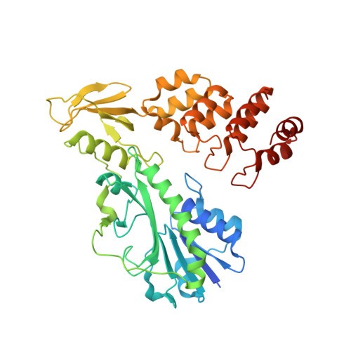
    N-Formylated sugars such as 3,6-dideoxy-3-formamido-d-glucose (Qui3NFo) have been observed on the lipopolysaccharides of various pathogenic bacteria, including Providencia alcalifaciens, a known cause of gastroenteritis. These unusual carbohydrates are synthesized in vivo as dTDP-linked sugars. The biosynthetic pathway for the production of dTDP-Qui3NFo requires five enzymes with the last step catalyzed by an N-formyltransferase that utilizes N(10)-tetrahydrofolate as a cofactor. Here we describe a structural and functional investigation of the P. alcalifaciens N-formyltransferase, hereafter referred to as QdtF. For this analysis, the structure of the dimeric enzyme was determined in the presence of N(5)-formyltetrahydrofolate, a stable cofactor, and dTDP-3,6-dideoxy-3-amino-d-glucose (dTDP-Qui3N) to 1.5 Å resolution. The overall fold of the subunit consists of three regions with the N-terminal and middle motifs followed by an ankyrin repeat domain. Whereas the ankyrin repeat is a common eukaryotic motif involved in protein-protein interactions, reports of its presence in prokaryotic enzymes have been limited. Unexpectedly, this ankyrin repeat houses a second binding pocket for dTDP-Qui3N, which is characterized by extensive interactions between the protein and the ligand. To address the effects of this second binding site on catalysis, a site-directed mutant protein, W305A, was constructed. Kinetic analyses demonstrated that the catalytic activity of the W305A variant was reduced by approximately 7-fold. The structure of the W305A mutant protein in complex with N(5)-formyltetrahydrofolate and dTDP-Qui3N was subsequently determined to 1.5 Å resolution. The electron density map clearly showed that ligand binding had been completely abolished in the auxiliary pocket. The wild-type enzyme was also tested for activity against dTDP-3,6-dideoxy-3-amino-d-galactose (dTDP-Fuc3N) as a substrate. Strikingly, sigmoidal kinetics indicating homotropic allosteric behavior were observed. Although the identity of the ligand that regulates QdtF activity in vivo is at present unknown, our results still provide the first example of an ankyrin repeat functioning in small molecule binding.


  • Organizational Affiliation

    Department of Biochemistry, University of Wisconsin , Madison, Wisconsin 53706, United States.


Macromolecules
Find similar proteins by:  (by identity cutoff)  |  3D Structure
Entity ID: 1
MoleculeChains Sequence LengthOrganismDetailsImage
TDP-3-aminoquinovose-N-formyltransferase417Providencia alcalifaciensMutation(s): 0 
Gene Names: qdtf
EC: 2.1.2.9
UniProt
Find proteins for F8RC03 (Providencia alcalifaciens)
Explore F8RC03 
Go to UniProtKB:  F8RC03
Entity Groups  
Sequence Clusters30% Identity50% Identity70% Identity90% Identity95% Identity100% Identity
UniProt GroupF8RC03
Sequence Annotations
Expand
  • Reference Sequence
Small Molecules
Ligands 6 Unique
IDChains Name / Formula / InChI Key2D Diagram3D Interactions
T3Q
Query on T3Q

Download Ideal Coordinates CCD File 
C [auth A],
D [auth A]
[(3R,4S,5S,6R)-4-amino-3,5-dihydroxy-6-methyloxan-2-yl][hydroxy-[[(2R,3S,5R)-3-hydroxy-5-(5-methyl-2,4-dioxopyrimidin-1-yl)oxolan-2-yl]methoxy]phosphoryl] hydrogen phosphate
C16 H27 N3 O14 P2
KVYJLJOGNUNRJK-HALQBZCBSA-N
FON
Query on FON

Download Ideal Coordinates CCD File 
B [auth A]N-{[4-({[(6R)-2-amino-5-formyl-4-oxo-1,4,5,6,7,8-hexahydropteridin-6-yl]methyl}amino)phenyl]carbonyl}-L-glutamic acid
C20 H23 N7 O7
VVIAGPKUTFNRDU-OLZOCXBDSA-N
EDO
Query on EDO

Download Ideal Coordinates CCD File 
L [auth A],
M [auth A],
N [auth A],
O [auth A],
P [auth A]
1,2-ETHANEDIOL
C2 H6 O2
LYCAIKOWRPUZTN-UHFFFAOYSA-N
K
Query on K

Download Ideal Coordinates CCD File 
H [auth A]POTASSIUM ION
K
NPYPAHLBTDXSSS-UHFFFAOYSA-N
CL
Query on CL

Download Ideal Coordinates CCD File 
K [auth A]CHLORIDE ION
Cl
VEXZGXHMUGYJMC-UHFFFAOYSA-M
NA
Query on NA

Download Ideal Coordinates CCD File 
E [auth A],
F [auth A],
G [auth A],
I [auth A],
J [auth A]
SODIUM ION
Na
FKNQFGJONOIPTF-UHFFFAOYSA-N
Experimental Data & Validation

Experimental Data

  • Method: X-RAY DIFFRACTION
  • Resolution: 1.50 Å
  • R-Value Free: 0.215 
  • R-Value Work: 0.183 
  • R-Value Observed: 0.185 
  • Space Group: P 41 21 2
Unit Cell:
Length ( Å )Angle ( ˚ )
a = 78.235α = 90
b = 78.235β = 90
c = 151.131γ = 90
Software Package:
Software NamePurpose
REFMACrefinement
SAINTdata reduction
SADABSdata scaling
PHASERphasing

Structure Validation

View Full Validation Report


Ligand Structure Quality Assessment 


Entry History 

Deposition Data

Revision History  (Full details and data files)

  • Version 1.0: 2015-01-21
    Type: Initial release
  • Version 1.1: 2015-02-04
    Changes: Database references
  • Version 1.2: 2015-02-11
    Changes: Database references
  • Version 1.3: 2023-09-27
    Changes: Data collection, Database references, Derived calculations, Refinement description, Source and taxonomy