4XBF | pdb_00004xbf

Structure of LSD1:CoREST in complex with ssRNA


Experimental Data Snapshot

  • Method: X-RAY DIFFRACTION
  • Resolution: 2.80 Å
  • R-Value Free: 
    0.227 (Depositor), 0.229 (DCC) 
  • R-Value Work: 
    0.201 (Depositor), 0.203 (DCC) 
  • R-Value Observed: 
    0.202 (Depositor) 

Starting Model: experimental
View more details

wwPDB Validation 3D Report Full Report

Validation slider image for 4XBF

Ligand Structure Quality Assessment 


This is version 1.2 of the entry. See complete history

Literature

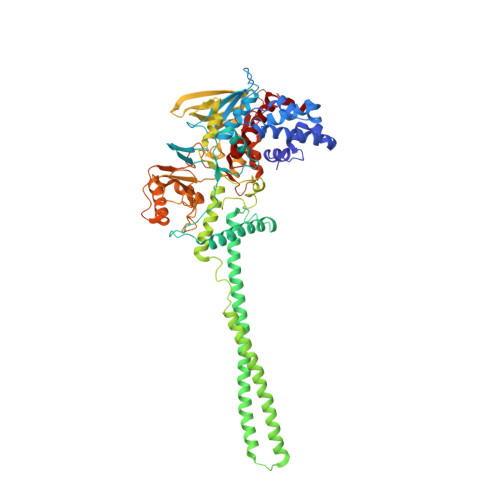
G-quadruplex RNA binding and recognition by the lysine-specific histone demethylase-1 enzyme.

Hirschi, A.Martin, W.J.Luka, Z.Loukachevitch, L.V.Reiter, N.J.

(2016) RNA 22: 1250-1260

  • DOI: https://doi.org/10.1261/rna.057265.116
  • Primary Citation Related Structures: 
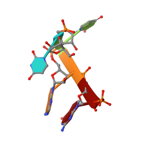
    4XBF

  • PubMed Abstract: 

    Lysine-specific histone demethylase 1 (LSD1) is an essential epigenetic regulator in metazoans and requires the co-repressor element-1 silencing transcription factor (CoREST) to efficiently catalyze the removal of mono- and dimethyl functional groups from histone 3 at lysine positions 4 and 9 (H3K4/9). LSD1 interacts with over 60 regulatory proteins and also associates with lncRNAs (TERRA, HOTAIR), suggesting a regulatory role for RNA in LSD1 function. We report that a stacked, intramolecular G-quadruplex (GQ) forming TERRA RNA (GG[UUAGGG]8UUA) binds tightly to the functional LSD1-CoREST complex (Kd ≈ 96 nM), in contrast to a single GQ RNA unit ([UUAGGG]4U), a GQ DNA ([TTAGGG]4T), or an unstructured single-stranded RNA. Stabilization of a parallel-stranded GQ RNA structure by monovalent potassium ions (K(+)) is required for high affinity binding to the LSD1-CoREST complex. These data indicate that LSD1 can distinguish between RNA and DNA as well as structured versus unstructured nucleotide motifs. Further, cross-linking mass spectrometry identified the primary location of GQ RNA binding within the SWIRM/amine oxidase domain (AOD) of LSD1. An ssRNA binding region adjacent to this GQ binding site was also identified via X-ray crystallography. This RNA binding interface is consistent with kinetic assays, demonstrating that a GQ-forming RNA can serve as a noncompetitive inhibitor of LSD1-catalyzed demethylation. The identification of a GQ RNA binding site coupled with kinetic data suggests that structured RNAs can function as regulatory molecules in LSD1-mediated mechanisms.


  • Organizational Affiliation
    • Department of Biochemistry, Vanderbilt University Medical Center, Nashville, Tennessee 37232-0146, USA.

Macromolecule Content 

  • Total Structure Weight: 92.39 kDa 
  • Atom Count: 6,486 
  • Modeled Residue Count: 804 
  • Deposited Residue Count: 804 
  • Unique protein chains: 2
  • Unique nucleic acid chains: 1

Macromolecules


Find similar proteins by:|  3D Structure
Entity ID: 1
MoleculeChains  Sequence LengthOrganismDetailsImage
Lysine-specific histone demethylase 1A666Homo sapiensMutation(s): 0 
Gene Names: KDM1AAOF2KDM1KIAA0601LSD1
EC: 1 (PDB Primary Data), 1.14.11 (UniProt), 1.14.99.66 (UniProt), 1.14.11.65 (UniProt)
UniProt & NIH Common Fund Data Resources
Find proteins for O60341 (Homo sapiens)
Explore O60341 
Go to UniProtKB:  O60341
PHAROS:  O60341
GTEx:  ENSG00000004487 
Entity Groups
Sequence Clusters30% Identity50% Identity70% Identity90% Identity95% Identity100% Identity
UniProt GroupO60341
Sequence Annotations
Expand
Reference Sequence
Find similar proteins by:|  3D Structure
Entity ID: 2
MoleculeChains  Sequence LengthOrganismDetailsImage
REST corepressor 1133Homo sapiensMutation(s): 0 
Gene Names: RCOR1KIAA0071RCOR
UniProt & NIH Common Fund Data Resources
Find proteins for Q9UKL0 (Homo sapiens)
Explore Q9UKL0 
Go to UniProtKB:  Q9UKL0
PHAROS:  Q9UKL0
GTEx:  ENSG00000089902 
Entity Groups
Sequence Clusters30% Identity50% Identity70% Identity90% Identity95% Identity100% Identity
UniProt GroupQ9UKL0
Sequence Annotations
Expand
Reference Sequence
Find similar nucleic acids by:  Sequence
Entity ID: 3
MoleculeChains LengthOrganismImage
RNA (5'-R(*UP*UP*AP*GP*G)-3')C [auth D]5Homo sapiens
Sequence Annotations
Expand
Reference Sequence

Small Molecules

Ligands 3 Unique
IDChains Name / Formula / InChI Key2D Diagram3D Interactions
FAD

Query on FAD


Download:Ideal Coordinates CCD File
D [auth A]FLAVIN-ADENINE DINUCLEOTIDE
C27 H33 N9 O15 P2
VWWQXMAJTJZDQX-UYBVJOGSSA-N
SO4

Query on SO4


Download:Ideal Coordinates CCD File
E [auth A],
F [auth A],
G [auth A],
H [auth A],
I [auth A]
SULFATE ION
O4 S
QAOWNCQODCNURD-UHFFFAOYSA-L
GOL

Query on GOL


Download:Ideal Coordinates CCD File
J [auth A]GLYCEROL
C3 H8 O3
PEDCQBHIVMGVHV-UHFFFAOYSA-N

Experimental Data & Validation

Experimental Data

  • Method: X-RAY DIFFRACTION
  • Resolution: 2.80 Å
  • R-Value Free:  0.227 (Depositor), 0.229 (DCC) 
  • R-Value Work:  0.201 (Depositor), 0.203 (DCC) 
  • R-Value Observed: 0.202 (Depositor) 
Space Group: I 2 2 2
Unit Cell:
Length ( Å )Angle ( ˚ )
a = 121.073α = 90
b = 178.411β = 90
c = 234.676γ = 90
Software Package:
Software NamePurpose
PHENIXrefinement
HKL-2000data reduction
Cootmodel building

Structure Validation

View Full Validation Report


Ligand Structure Quality Assessment 


Entry History 

& Funding Information

Deposition Data


Funding OrganizationLocationGrant Number
American Cancer SocietyUnited StatesIRG-58-009-54

Revision History  (Full details and data files)

  • Version 1.0: 2016-04-27
    Type: Initial release
  • Version 1.1: 2017-05-03
    Changes: Data collection, Database references
  • Version 1.2: 2023-09-27
    Changes: Data collection, Database references, Refinement description