4XB3 | pdb_00004xb3

Structure of dextran glucosidase


Experimental Data Snapshot

  • Method: X-RAY DIFFRACTION
  • Resolution: 2.09 Å
  • R-Value Free: 
    0.219 (Depositor), 0.221 (DCC) 
  • R-Value Work: 
    0.179 (Depositor), 0.183 (DCC) 
  • R-Value Observed: 
    0.181 (Depositor) 

wwPDB Validation 3D Report Full Report

Validation slider image for 4XB3

This is version 1.2 of the entry. See complete history

Literature

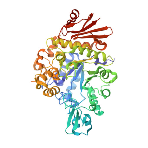
Structural insights into the catalytic reaction that is involved in the reorientation of Trp238 at the substrate-binding site in GH13 dextran glucosidase

Kobayashi, M.Saburi, W.Nakatsuka, D.Hondoh, H.Kato, K.Okuyama, M.Mori, H.Kimura, A.Yao, M.

(2015) FEBS Lett 589: 484-489

  • DOI: https://doi.org/10.1016/j.febslet.2015.01.005
  • Primary Citation Related Structures: 
    4WLC, 4XB3

  • PubMed Abstract: 

    Streptococcus mutans dextran glucosidase (SmDG) belongs to glycoside hydrolase family 13, and catalyzes both the hydrolysis of substrates such as isomaltooligosaccharides and subsequent transglucosylation to form α-(1→6)-glucosidic linkage at the substrate non-reducing ends. Here, we report the 2.4Å resolution crystal structure of glucosyl-enzyme intermediate of SmDG. In the obtained structure, the Trp238 side-chain that constitutes the substrate-binding site turned away from the active pocket, concurrently with conformational changes of the nucleophile and the acid/base residues. Different conformations of Trp238 in each reaction stage indicated its flexibility. Considering the results of kinetic analyses, such flexibility may reflect a requirement for the reaction mechanism of SmDG.


  • Organizational Affiliation
    • Graduate School of Life Science, Hokkaido University, Kita-10, Nishi-8, Kita-ku, Sapporo 060-0810, Japan.

Macromolecule Content 

  • Total Structure Weight: 62.51 kDa 
  • Atom Count: 4,749 
  • Modeled Residue Count: 536 
  • Deposited Residue Count: 536 
  • Unique protein chains: 1

Macromolecules

Find similar proteins by:|  3D Structure
Entity ID: 1
MoleculeChains  Sequence LengthOrganismDetailsImage
Glucan 1,6-alpha-glucosidase536Streptococcus mutans UA159Mutation(s): 2 
Gene Names: dexBSMU_883
EC: 3.2.1.70
UniProt
Find proteins for Q99040 (Streptococcus mutans serotype c (strain ATCC 700610 / UA159))
Explore Q99040 
Go to UniProtKB:  Q99040
Entity Groups
Sequence Clusters30% Identity50% Identity70% Identity90% Identity95% Identity100% Identity
UniProt GroupQ99040
Sequence Annotations
Expand
Reference Sequence

Experimental Data & Validation

Experimental Data

  • Method: X-RAY DIFFRACTION
  • Resolution: 2.09 Å
  • R-Value Free:  0.219 (Depositor), 0.221 (DCC) 
  • R-Value Work:  0.179 (Depositor), 0.183 (DCC) 
  • R-Value Observed: 0.181 (Depositor) 
Space Group: P 21 21 21
Unit Cell:
Length ( Å )Angle ( ˚ )
a = 72.151α = 90
b = 82.55β = 90
c = 103.559γ = 90
Software Package:
Software NamePurpose
PHENIXrefinement

Structure Validation

View Full Validation Report


Entry History 

& Funding Information

Deposition Data


Funding OrganizationLocationGrant Number
the Japan Society for the Promotion of ScienceJapan24-871
the Ministry of Education, Culture, Sports, Science and TechnologyJapan--

Revision History  (Full details and data files)

  • Version 1.0: 2015-08-26
    Type: Initial release
  • Version 1.1: 2020-02-05
    Changes: Data collection, Database references, Derived calculations
  • Version 1.2: 2024-03-20
    Changes: Data collection, Database references, Derived calculations