4X9J | pdb_00004x9j

EGR-1 with Doubly Methylated DNA


Experimental Data Snapshot

  • Method: X-RAY DIFFRACTION
  • Resolution: 1.41 Å
  • R-Value Free: 
    0.200 (Depositor), 0.202 (DCC) 
  • R-Value Work: 
    0.172 (Depositor), 0.176 (DCC) 
  • R-Value Observed: 
    0.174 (Depositor) 

Starting Model: experimental
View more details

wwPDB Validation 3D Report Full Report

Validation slider image for 4X9J

This is version 1.4 of the entry. See complete history

Literature

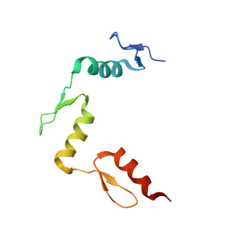
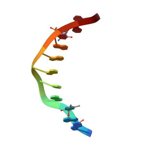
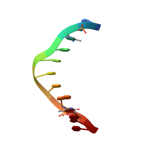
Structural impact of complete CpG methylation within target DNA on specific complex formation of the inducible transcription factor Egr-1.

Zandarashvili, L.White, M.A.Esadze, A.Iwahara, J.

(2015) FEBS Lett 589: 1748-1753

  • DOI: https://doi.org/10.1016/j.febslet.2015.05.022
  • Primary Citation Related Structures: 
    4X9J

  • PubMed Abstract: 

    The inducible transcription factor Egr-1 binds specifically to 9-bp target sequences containing two CpG sites that can potentially be methylated at four cytosine bases. Although it appears that complete CpG methylation would make an unfavorable steric clash in the previous crystal structures of the complexes with unmethylated or partially methylated DNA, our affinity data suggest that DNA recognition by Egr-1 is insensitive to CpG methylation. We have determined, at a 1.4-Å resolution, the crystal structure of the Egr-1 zinc-finger complex with completely methylated target DNA. Structural comparison of the three different methylation states reveals why Egr-1 can recognize the target sequences regardless of CpG methylation.


  • Organizational Affiliation
    • Department of Biochemistry & Molecular Biology, Sealy Center for Structural Biology and Molecular Biophysics, University of Texas Medical Branch, Galveston, TX 77555-1068, USA.

Macromolecule Content 

  • Total Structure Weight: 17.68 kDa 
  • Atom Count: 1,419 
  • Modeled Residue Count: 108 
  • Deposited Residue Count: 111 
  • Unique protein chains: 1
  • Unique nucleic acid chains: 2

Macromolecules


Find similar proteins by:|  3D Structure
Entity ID: 1
MoleculeChains  Sequence LengthOrganismDetailsImage
Early growth response protein 189Homo sapiensMutation(s): 0 
Gene Names: EGR1KROX24ZNF225
UniProt & NIH Common Fund Data Resources
Find proteins for P18146 (Homo sapiens)
Explore P18146 
Go to UniProtKB:  P18146
PHAROS:  P18146
GTEx:  ENSG00000120738 
Entity Groups
Sequence Clusters30% Identity50% Identity70% Identity90% Identity95% Identity100% Identity
UniProt GroupP18146
Sequence Annotations
Expand
Reference Sequence
Find similar nucleic acids by:  Sequence
Entity ID: 2
MoleculeChains LengthOrganismImage
DNA (5'-D(*AP*GP*(5CM)P*GP*TP*GP*GP*GP*(5CM)P*GP*T)-3')11synthetic construct
Sequence Annotations
Expand
Reference Sequence
Find similar nucleic acids by:  Sequence
Entity ID: 3
MoleculeChains LengthOrganismImage
DNA (5'-D(*TP*AP*(5CM)P*GP*CP*CP*CP*AP*(5CM)P*GP*C)-3')11synthetic construct
Sequence Annotations
Expand
Reference Sequence

Experimental Data & Validation

Experimental Data

  • Method: X-RAY DIFFRACTION
  • Resolution: 1.41 Å
  • R-Value Free:  0.200 (Depositor), 0.202 (DCC) 
  • R-Value Work:  0.172 (Depositor), 0.176 (DCC) 
  • R-Value Observed: 0.174 (Depositor) 
Space Group: C 2 2 21
Unit Cell:
Length ( Å )Angle ( ˚ )
a = 44.009α = 90
b = 55.989β = 90
c = 128.952γ = 90
Software Package:
Software NamePurpose
PHENIXrefinement
HKL-2000data reduction
PDB_EXTRACTdata extraction
HKL-2000data scaling
PHENIXphasing

Structure Validation

View Full Validation Report


Entry History 

Deposition Data

Revision History  (Full details and data files)

  • Version 1.0: 2015-05-13
    Type: Initial release
  • Version 1.1: 2015-06-03
    Changes: Database references
  • Version 1.2: 2015-07-15
    Changes: Database references
  • Version 1.3: 2017-09-27
    Changes: Author supporting evidence, Database references, Derived calculations, Source and taxonomy
  • Version 1.4: 2023-09-27
    Changes: Data collection, Database references, Derived calculations, Refinement description