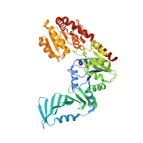
4X7P

Crystal structure of apo S. aureus TarM


Experimental Data Snapshot

  • Method: X-RAY DIFFRACTION
  • Resolution: 3.40 Å
  • R-Value Free: 0.187 
  • R-Value Work: 0.173 
  • R-Value Observed: 0.173 

wwPDB Validation   3D Report Full Report


This is version 1.5 of the entry. See complete history


Literature

Structure and mechanism of Staphylococcus aureus TarM, the wall teichoic acid alpha-glycosyltransferase.

Sobhanifar, S.Worrall, L.J.Gruninger, R.J.Wasney, G.A.Blaukopf, M.Baumann, L.Lameignere, E.Solomonson, M.Brown, E.D.Withers, S.G.Strynadka, N.C.

(2015) Proc Natl Acad Sci U S A 112: E576-E585

  • DOI: https://doi.org/10.1073/pnas.1418084112
  • Primary Citation of Related Structures:  
    4X6L, 4X7M, 4X7P, 4X7R

  • PubMed Abstract: 

    Unique to Gram-positive bacteria, wall teichoic acids are anionic glycopolymers cross-stitched to a thick layer of peptidoglycan. The polyol phosphate subunits of these glycopolymers are decorated with GlcNAc sugars that are involved in phage binding, genetic exchange, host antibody response, resistance, and virulence. The search for the enzymes responsible for GlcNAcylation in Staphylococcus aureus has recently identified TarM and TarS with respective α- and β-(1-4) glycosyltransferase activities. The stereochemistry of the GlcNAc attachment is important in balancing biological processes, such that the interplay of TarM and TarS is likely important for bacterial pathogenicity and survival. Here we present the crystal structure of TarM in an unusual ternary-like complex consisting of a polymeric acceptor substrate analog, UDP from a hydrolyzed donor, and an α-glyceryl-GlcNAc product formed in situ. These structures support an internal nucleophilic substitution-like mechanism, lend new mechanistic insight into the glycosylation of glycopolymers, and reveal a trimerization domain with a likely role in acceptor substrate scaffolding.


  • Organizational Affiliation

    Department of Biochemistry and Center for Blood Research, University of British Columbia, Vancouver, BC, Canada V6T 1Z3;


Macromolecules
Find similar proteins by:  (by identity cutoff)  |  3D Structure
Entity ID: 1
MoleculeChains Sequence LengthOrganismDetailsImage
TarM
A, B
493Staphylococcus aureus subsp. aureus 21178Mutation(s): 0 
Gene Names: SA21178_0837
UniProt
Find proteins for A0A0H2WWV6 (Staphylococcus aureus (strain COL))
Explore A0A0H2WWV6 
Go to UniProtKB:  A0A0H2WWV6
Entity Groups  
Sequence Clusters30% Identity50% Identity70% Identity90% Identity95% Identity100% Identity
UniProt GroupA0A0H2WWV6
Sequence Annotations
Expand
  • Reference Sequence
Small Molecules
Ligands 1 Unique
IDChains Name / Formula / InChI Key2D Diagram3D Interactions
SO4
Query on SO4

Download Ideal Coordinates CCD File 
C [auth A]
D [auth A]
E [auth A]
F [auth A]
G [auth A]
C [auth A],
D [auth A],
E [auth A],
F [auth A],
G [auth A],
H [auth A],
I [auth A],
J [auth A],
K [auth B],
L [auth B],
M [auth B],
N [auth B],
O [auth B],
P [auth B],
Q [auth B],
R [auth B]
SULFATE ION
O4 S
QAOWNCQODCNURD-UHFFFAOYSA-L
Experimental Data & Validation

Experimental Data

  • Method: X-RAY DIFFRACTION
  • Resolution: 3.40 Å
  • R-Value Free: 0.187 
  • R-Value Work: 0.173 
  • R-Value Observed: 0.173 
  • Space Group: P 63
Unit Cell:
Length ( Å )Angle ( ˚ )
a = 208.599α = 90
b = 208.599β = 90
c = 120.751γ = 120
Software Package:
Software NamePurpose
xia2data scaling
SCALAdata scaling
PHASERphasing
BUSTERrefinement
REFMACrefinement
PDB_EXTRACTdata extraction

Structure Validation

View Full Validation Report


Entry History & Funding Information

Deposition Data


Funding OrganizationLocationGrant Number
Canadian Institutes of Health Research (CIHR)Canada--
Howard Hughes Medical Institute (HHMI)United States--

Revision History  (Full details and data files)

  • Version 1.0: 2015-02-18
    Type: Initial release
  • Version 1.1: 2015-02-25
    Changes: Database references
  • Version 1.2: 2017-09-13
    Changes: Author supporting evidence, Database references, Derived calculations, Source and taxonomy
  • Version 1.3: 2017-11-22
    Changes: Refinement description
  • Version 1.4: 2019-11-20
    Changes: Author supporting evidence
  • Version 1.5: 2024-02-28
    Changes: Data collection, Database references