4X5T | pdb_00004x5t

alpha 1 glycine receptor transmembrane structure fused to the extracellular domain of GLIC


Experimental Data Snapshot

  • Method: X-RAY DIFFRACTION
  • Resolution: 3.50 Å
  • R-Value Free: 
    0.270 (Depositor), 0.304 (DCC) 
  • R-Value Work: 
    0.254 (Depositor), 0.277 (DCC) 
  • R-Value Observed: 
    0.254 (Depositor) 

Starting Model: experimental
View more details

wwPDB Validation 3D Report Full Report

Validation slider image for 4X5T

This is version 1.3 of the entry. See complete history

Literature

Allosteric and hyperekplexic mutant phenotypes investigated on an alpha 1 glycine receptor transmembrane structure.

Moraga-Cid, G.Sauguet, L.Huon, C.Malherbe, L.Girard-Blanc, C.Petres, S.Murail, S.Taly, A.Baaden, M.Delarue, M.Corringer, P.J.

(2015) Proc Natl Acad Sci U S A 112: 2865-2870

  • DOI: https://doi.org/10.1073/pnas.1417864112
  • Primary Citation Related Structures: 
    4X5T

  • PubMed Abstract: 

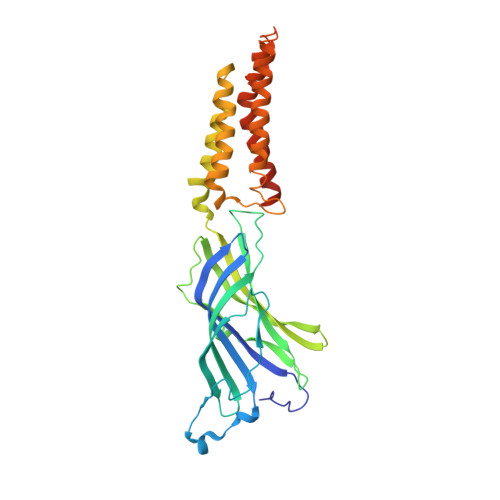
    The glycine receptor (GlyR) is a pentameric ligand-gated ion channel (pLGIC) mediating inhibitory transmission in the nervous system. Its transmembrane domain (TMD) is the target of allosteric modulators such as general anesthetics and ethanol and is a major locus for hyperekplexic congenital mutations altering the allosteric transitions of activation or desensitization. We previously showed that the TMD of the human α1GlyR could be fused to the extracellular domain of GLIC, a bacterial pLGIC, to form a functional chimera called Lily. Here, we overexpress Lily in Schneider 2 insect cells and solve its structure by X-ray crystallography at 3.5 Å resolution. The TMD of the α1GlyR adopts a closed-channel conformation involving a single ring of hydrophobic residues at the center of the pore. Electrophysiological recordings show that the phenotypes of key allosteric mutations of the α1GlyR, scattered all along the pore, are qualitatively preserved in this chimera, including those that confer decreased sensitivity to agonists, constitutive activity, decreased activation kinetics, or increased desensitization kinetics. Combined structural and functional data indicate a pore-opening mechanism for the α1GlyR, suggesting a structural explanation for the effect of some key hyperekplexic allosteric mutations. The first X-ray structure of the TMD of the α1GlyR solved here using GLIC as a scaffold paves the way for mechanistic investigation and design of allosteric modulators of a human receptor.


  • Organizational Affiliation
    • Channel-Receptors Unit, Institut Pasteur, 75015 Paris, France; CNRS UMR 3571, 75015 Paris, France;

Macromolecule Content 

  • Total Structure Weight: 190.26 kDa 
  • Atom Count: 11,485 
  • Modeled Residue Count: 1,536 
  • Deposited Residue Count: 1,650 
  • Unique protein chains: 1

Macromolecules

Find similar proteins by:|  3D Structure
Entity ID: 1
MoleculeChains  Sequence LengthOrganismDetailsImage
Proton-gated ion channel,GLRA1 protein,GLRA1 protein
A, B, C, D, E
330Gloeobacter violaceus PCC 7421Homo sapiensMutation(s): 0 
Gene Names: glvIglr4197GLRA1
Membrane Entity: Yes 
UniProt & NIH Common Fund Data Resources
Find proteins for P23415 (Homo sapiens)
Explore P23415 
Go to UniProtKB:  P23415
PHAROS:  P23415
GTEx:  ENSG00000145888 
Find proteins for Q7NDN8 (Gloeobacter violaceus (strain ATCC 29082 / PCC 7421))
Explore Q7NDN8 
Go to UniProtKB:  Q7NDN8
Entity Groups
Sequence Clusters30% Identity50% Identity70% Identity90% Identity95% Identity100% Identity
UniProt GroupsQ7NDN8P23415
Sequence Annotations
Expand
Reference Sequence

Small Molecules

Ligands 3 Unique
IDChains Name / Formula / InChI Key2D Diagram3D Interactions
ACT

Query on ACT


Download:Ideal Coordinates CCD File
H [auth A],
L [auth B],
P [auth C],
S [auth D],
V [auth E]
ACETATE ION
C2 H3 O2
QTBSBXVTEAMEQO-UHFFFAOYSA-M
NI

Query on NI


Download:Ideal Coordinates CCD File
F [auth A]
I [auth B]
M [auth C]
Q [auth D]
R [auth D]
F [auth A],
I [auth B],
M [auth C],
Q [auth D],
R [auth D],
T [auth E]
NICKEL (II) ION
Ni
VEQPNABPJHWNSG-UHFFFAOYSA-N
CL

Query on CL


Download:Ideal Coordinates CCD File
G [auth A]
J [auth B]
K [auth B]
N [auth C]
O [auth C]
G [auth A],
J [auth B],
K [auth B],
N [auth C],
O [auth C],
U [auth E]
CHLORIDE ION
Cl
VEXZGXHMUGYJMC-UHFFFAOYSA-M

Experimental Data & Validation

Experimental Data

  • Method: X-RAY DIFFRACTION
  • Resolution: 3.50 Å
  • R-Value Free:  0.270 (Depositor), 0.304 (DCC) 
  • R-Value Work:  0.254 (Depositor), 0.277 (DCC) 
  • R-Value Observed: 0.254 (Depositor) 
Space Group: P 21 21 21
Unit Cell:
Length ( Å )Angle ( ˚ )
a = 118.894α = 90
b = 132.304β = 90
c = 190.517γ = 90
Software Package:
Software NamePurpose
BUSTERrefinement
XDSdata reduction
SCALAdata scaling
PHASERphasing

Structure Validation

View Full Validation Report


Entry History 

Deposition Data

Revision History  (Full details and data files)

  • Version 1.0: 2015-02-25
    Type: Initial release
  • Version 1.1: 2015-03-11
    Changes: Database references
  • Version 1.2: 2015-03-18
    Changes: Database references
  • Version 1.3: 2024-01-10
    Changes: Data collection, Database references, Refinement description