


Crystal structure of an oxidoreductase/ short chain dehydrogenase from Brucella ovis
Lukacs, C.M., Edwards, T.E., Lorimer, D.D.To be published.
Experimental Data Snapshot
wwPDB Validation 3D Report Full Report

Entity ID: 1 | |||||
|---|---|---|---|---|---|
| Molecule | Chains | Sequence Length | Organism | Details | Image |
| Oxidoreductase, short chain dehydrogenase/reductase family | 276 | Brucella ovis ATCC 25840 | Mutation(s): 0 Gene Names: BOV_0113 | 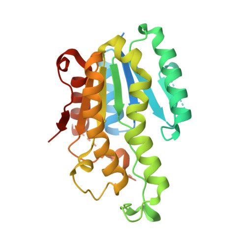 | |
UniProt | |||||
Find proteins for A0A0H3AQ75 (Brucella ovis (strain ATCC 25840 / 63/290 / NCTC 10512)) Explore A0A0H3AQ75 Go to UniProtKB: A0A0H3AQ75 | |||||
Entity Groups | |||||
| Sequence Clusters | 30% Identity50% Identity70% Identity90% Identity95% Identity100% Identity | ||||
| UniProt Group | A0A0H3AQ75 | ||||
Sequence AnnotationsExpand | |||||
Reference Sequence | |||||
| Ligands 1 Unique | |||||
|---|---|---|---|---|---|
| ID | Chains | Name / Formula / InChI Key | 2D Diagram | 3D Interactions | |
| MPD Download:Ideal Coordinates CCD File | B [auth A], C [auth A] | (4S)-2-METHYL-2,4-PENTANEDIOL C6 H14 O2 SVTBMSDMJJWYQN-YFKPBYRVSA-N |  | ||
| Length ( Å ) | Angle ( ˚ ) |
|---|---|
| a = 53.19 | α = 90 |
| b = 100.88 | β = 90 |
| c = 101.41 | γ = 90 |
| Software Name | Purpose |
|---|---|
| PHENIX | refinement |
| XDS | data reduction |
| XSCALE | data scaling |
| PHASER | phasing |


