4X25

Structural basis for mutation-induced destabilization of Profilin 1 in ALS


Experimental Data Snapshot

  • Method: X-RAY DIFFRACTION
  • Resolution: 2.23 Å
  • R-Value Free: 0.238 
  • R-Value Work: 0.195 
  • R-Value Observed: 0.198 

wwPDB Validation   3D Report Full Report


This is version 1.4 of the entry. See complete history


Literature

Structural basis for mutation-induced destabilization of profilin 1 in ALS.

Boopathy, S.Silvas, T.V.Tischbein, M.Jansen, S.Shandilya, S.M.Zitzewitz, J.A.Landers, J.E.Goode, B.L.Schiffer, C.A.Bosco, D.A.

(2015) Proc Natl Acad Sci U S A 112: 7984-7989

  • DOI: https://doi.org/10.1073/pnas.1424108112
  • Primary Citation of Related Structures:  
    4X1L, 4X1M, 4X25

  • PubMed Abstract: 

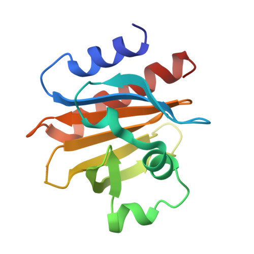
    Mutations in profilin 1 (PFN1) are associated with amyotrophic lateral sclerosis (ALS); however, the pathological mechanism of PFN1 in this fatal disease is unknown. We demonstrate that ALS-linked mutations severely destabilize the native conformation of PFN1 in vitro and cause accelerated turnover of the PFN1 protein in cells. This mutation-induced destabilization can account for the high propensity of ALS-linked variants to aggregate and also provides rationale for their reported loss-of-function phenotypes in cell-based assays. The source of this destabilization is illuminated by the X-ray crystal structures of several PFN1 proteins, revealing an expanded cavity near the protein core of the destabilized M114T variant. In contrast, the E117G mutation only modestly perturbs the structure and stability of PFN1, an observation that reconciles the occurrence of this mutation in the control population. These findings suggest that a destabilized form of PFN1 underlies PFN1-mediated ALS pathogenesis.


  • Organizational Affiliation

    Department of Neurology, University of Massachusetts Medical School, Worcester, MA 01605;


Macromolecules
Find similar proteins by:  (by identity cutoff)  |  3D Structure
Entity ID: 1
MoleculeChains Sequence LengthOrganismDetailsImage
Profilin-1
A, B
140Homo sapiensMutation(s): 1 
Gene Names: PFN1
UniProt & NIH Common Fund Data Resources
Find proteins for P07737 (Homo sapiens)
Explore P07737 
Go to UniProtKB:  P07737
PHAROS:  P07737
GTEx:  ENSG00000108518 
Entity Groups  
Sequence Clusters30% Identity50% Identity70% Identity90% Identity95% Identity100% Identity
UniProt GroupP07737
Sequence Annotations
Expand
  • Reference Sequence
Experimental Data & Validation

Experimental Data

  • Method: X-RAY DIFFRACTION
  • Resolution: 2.23 Å
  • R-Value Free: 0.238 
  • R-Value Work: 0.195 
  • R-Value Observed: 0.198 
  • Space Group: P 6
Unit Cell:
Length ( Å )Angle ( ˚ )
a = 81.69α = 90
b = 81.69β = 90
c = 65.35γ = 120
Software Package:
Software NamePurpose
HKL-3000data collection
xia2data scaling
XDSdata reduction
XSCALEdata scaling
Cootmodel building
PHENIXrefinement
PHASERphasing

Structure Validation

View Full Validation Report


Entry History 

Deposition Data

Revision History  (Full details and data files)

  • Version 1.0: 2015-06-10
    Type: Initial release
  • Version 1.1: 2015-06-17
    Changes: Database references, Structure summary
  • Version 1.2: 2017-11-22
    Changes: Derived calculations, Refinement description, Source and taxonomy
  • Version 1.3: 2018-04-25
    Changes: Data collection, Database references
  • Version 1.4: 2024-02-28
    Changes: Data collection, Database references