4X20 | pdb_00004x20

Discovery of cytotoxic Dolastatin 10 analogs with N-terminal modifications


Experimental Data Snapshot

  • Method: X-RAY DIFFRACTION
  • Resolution: 3.50 Å
  • R-Value Free: 
    0.252 (Depositor), 0.270 (DCC) 
  • R-Value Work: 
    0.181 (Depositor), 0.200 (DCC) 
  • R-Value Observed: 
    0.184 (Depositor) 

Starting Model: experimental
View more details

wwPDB Validation 3D Report Full Report

Validation slider image for 4X20

Ligand Structure Quality Assessment 


This is version 1.2 of the entry. See complete history

Literature

Discovery of cytotoxic dolastatin 10 analogues with N-terminal modifications.

Maderna, A.Doroski, M.Subramanyam, C.Porte, A.Leverett, C.A.Vetelino, B.C.Chen, Z.Risley, H.Parris, K.Pandit, J.Varghese, A.H.Shanker, S.Song, C.Sukuru, S.C.Farley, K.A.Wagenaar, M.M.Shapiro, M.J.Musto, S.Lam, M.H.Loganzo, F.O'Donnell, C.J.

(2014) J Med Chem 57: 10527-10543

  • DOI: https://doi.org/10.1021/jm501649k
  • Primary Citation Related Structures: 
    4X1I, 4X1K, 4X1Y, 4X20

  • PubMed Abstract: 

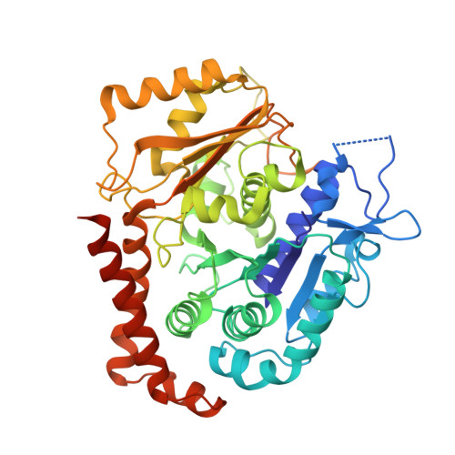
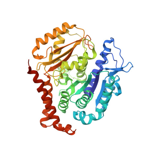
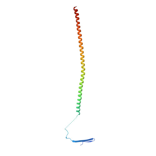
    Auristatins, synthetic analogues of the antineoplastic natural product Dolastatin 10, are ultrapotent cytotoxic microtubule inhibitors that are clinically used as payloads in antibody-drug conjugates (ADCs). The design and synthesis of several new auristatin analogues with N-terminal modifications that include amino acids with α,α-disubstituted carbon atoms are described, including the discovery of our lead auristatin, PF-06380101. This modification of the peptide structure is unprecedented and led to analogues with excellent potencies in tumor cell proliferation assays and differential ADME properties when compared to other synthetic auristatin analogues that are used in the preparation of ADCs. In addition, auristatin cocrystal structures with tubulin are being presented that allow for the detailed examination of their binding modes. A surprising finding is that all analyzed analogues have a cis-configuration at the Val-Dil amide bond in their functionally relevant tubulin bound state, whereas in solution this bond is exclusively in the trans-configuration. This remarkable observation shines light onto the preferred binding mode of auristatins and serves as a valuable tool for structure-based drug design.


  • Organizational Affiliation
    • Worldwide Medicinal Chemistry, Oncology, Pfizer Worldwide Research and Development , Eastern Point Road, Groton, Connecticut 06340, United States.

Macromolecule Content 

  • Total Structure Weight: 221.28 kDa 
  • Atom Count: 14,717 
  • Modeled Residue Count: 1,840 
  • Deposited Residue Count: 1,934 
  • Unique protein chains: 3

Macromolecules

Find similar proteins by:|  3D Structure
Entity ID: 1
MoleculeChains  Sequence LengthOrganismDetailsImage
Tubulin alpha chain
A, C
451Ovis ariesMutation(s): 0 
UniProt
Find proteins for D0VWZ0 (Ovis aries)
Explore D0VWZ0 
Go to UniProtKB:  D0VWZ0
Entity Groups
Sequence Clusters30% Identity50% Identity70% Identity90% Identity95% Identity100% Identity
UniProt GroupD0VWZ0
Sequence Annotations
Expand
Reference Sequence
Find similar proteins by:|  3D Structure
Entity ID: 2
MoleculeChains  Sequence LengthOrganismDetailsImage
Tubulin beta chain
B, D
445Ovis ariesMutation(s): 0 
UniProt
Find proteins for D0VWY9 (Ovis aries)
Explore D0VWY9 
Go to UniProtKB:  D0VWY9
Entity Groups
Sequence Clusters30% Identity50% Identity70% Identity90% Identity95% Identity100% Identity
UniProt GroupD0VWY9
Sequence Annotations
Expand
Reference Sequence
Find similar proteins by:|  3D Structure
Entity ID: 3
MoleculeChains  Sequence LengthOrganismDetailsImage
Stathmin-4142Rattus norvegicusMutation(s): 2 
Gene Names: Stmn4
UniProt
Find proteins for P63043 (Rattus norvegicus)
Explore P63043 
Go to UniProtKB:  P63043
Entity Groups
Sequence Clusters30% Identity50% Identity70% Identity90% Identity95% Identity100% Identity
UniProt GroupP63043
Sequence Annotations
Expand
Reference Sequence

Small Molecules

Ligands 5 Unique
IDChains Name / Formula / InChI Key2D Diagram3D Interactions
3WY

Query on 3WY


Download:Ideal Coordinates CCD File
J [auth B],
O [auth D]
2-methyl-L-prolyl-N-[(3R,4S,5S)-1-{(2S)-2-[(1R,2R)-3-{[(1S)-1-carboxy-2-phenylethyl]amino}-1-methoxy-2-methyl-3-oxopropyl]pyrrolidin-1-yl}-3-methoxy-5-methyl-1-oxoheptan-4-yl]-N-methyl-L-valinamide
C39 H63 N5 O8
IHPVMHRTGNEZNG-SJZZQOMKSA-N
GTP

Query on GTP


Download:Ideal Coordinates CCD File
F [auth A],
K [auth C]
GUANOSINE-5'-TRIPHOSPHATE
C10 H16 N5 O14 P3
XKMLYUALXHKNFT-UUOKFMHZSA-N
GDP

Query on GDP


Download:Ideal Coordinates CCD File
H [auth B],
M [auth D]
GUANOSINE-5'-DIPHOSPHATE
C10 H15 N5 O11 P2
QGWNDRXFNXRZMB-UUOKFMHZSA-N
LOC

Query on LOC


Download:Ideal Coordinates CCD File
I [auth B],
N [auth D]
N-[(7S)-1,2,3,10-tetramethoxy-9-oxo-6,7-dihydro-5H-benzo[d]heptalen-7-yl]ethanamide
C22 H25 N O6
IAKHMKGGTNLKSZ-INIZCTEOSA-N
MG

Query on MG


Download:Ideal Coordinates CCD File
G [auth A],
L [auth C]
MAGNESIUM ION
Mg
JLVVSXFLKOJNIY-UHFFFAOYSA-N

Experimental Data & Validation

Experimental Data

  • Method: X-RAY DIFFRACTION
  • Resolution: 3.50 Å
  • R-Value Free:  0.252 (Depositor), 0.270 (DCC) 
  • R-Value Work:  0.181 (Depositor), 0.200 (DCC) 
  • R-Value Observed: 0.184 (Depositor) 
Space Group: P 21 21 21
Unit Cell:
Length ( Å )Angle ( ˚ )
a = 66.24α = 90
b = 128.27β = 90
c = 254.14γ = 90
Software Package:
Software NamePurpose
BUSTERrefinement
DENZOdata reduction
PDB_EXTRACTdata extraction
SCALEPACKdata scaling
BUSTERphasing

Structure Validation

View Full Validation Report


Ligand Structure Quality Assessment 


Entry History 

Deposition Data

  • Released Date: 2015-03-25 
  • Deposition Author(s): Parris, K.D.

Revision History  (Full details and data files)

  • Version 1.0: 2015-03-25
    Type: Initial release
  • Version 1.1: 2015-04-08
    Changes: Non-polymer description
  • Version 1.2: 2023-09-27
    Changes: Data collection, Database references, Derived calculations, Refinement description, Source and taxonomy