4WZV | pdb_00004wzv

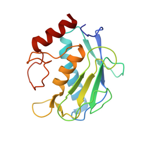
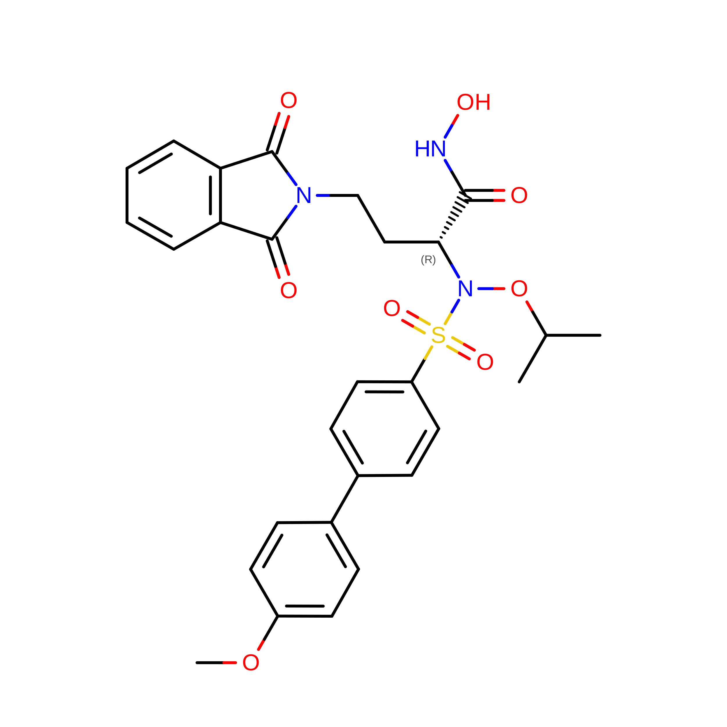
Crystal structure of a hydroxamate based inhibitor EN140 in complex with the MMP-9 catalytic domain


Experimental Data Snapshot

  • Method: X-RAY DIFFRACTION
  • Resolution: 1.65 Å
  • R-Value Free: 
    0.230 (Depositor), 0.230 (DCC) 
  • R-Value Work: 
    0.176 (Depositor), 0.177 (DCC) 
  • R-Value Observed: 
    0.179 (Depositor) 

Starting Model: experimental
View more details

wwPDB Validation 3D Report Full Report

Validation slider image for 4WZV

Ligand Structure Quality Assessment 


This is version 1.2 of the entry. See complete history. 

Literature

N-O-Isopropyl Sulfonamido-Based Hydroxamates as Matrix Metalloproteinase Inhibitors: Hit Selection and in Vivo Antiangiogenic Activity.

Nuti, E., Cantelmo, A.R., Gallo, C., Bruno, A., Bassani, B., Camodeca, C., Tuccinardi, T., Vera, L., Orlandini, E., Nencetti, S., Stura, E.A., Martinelli, A., Dive, V., Albini, A., Rossello, A.

(2015) J Med Chem 58: 7224-7240

  • DOI: https://doi.org/10.1021/acs.jmedchem.5b00367
  • Primary Citation Related Structures: 
    4WZV, 4XCT

  • PubMed Abstract: 

    Matrix metalloproteinases (MMPs) have been shown to be involved in tumor-induced angiogenesis. In particular, MMP-2, MMP-9, and MMP-14 have been reported to be crucial for tumor angiogenesis and the formation of metastasis, thus becoming attractive targets in cancer therapy. Here, we report our optimization effort to identify novel N-isopropoxy-arylsulfonamide hydroxamates with improved inhibitory activity toward MMP-2, MMP-9, and MMP-14 with respect to the previously discovered compound 1. A new series of hydroxamates was designed, synthesized, and tested for their antiangiogenic activity using in vitro assays with human umbilical vein endothelial cells (HUVECs). A nanomolar MMP-2, MMP-9, and MMP-14 inhibitor was identified, compound 3, able to potently inhibit angiogenesis in vitro and also in vivo in the matrigel sponge assay in mice. Finally, X-ray crystallographic and docking studies were conducted for compound 3 in order to investigate its binding mode to MMP-9 and MMP-14.


  • Organizational Affiliation: 
    • Dipartimento di Farmacia, Università di Pisa , via Bonanno 6, 56126 Pisa, Italy.

Macromolecule Content 

  • Total Structure Weight: 38.44 kDa 
  • Atom Count: 3,442 
  • Modeled Residue Count: 316 
  • Deposited Residue Count: 320 
  • Unique protein chains: 1

Macromolecules

Find similar proteins by:|  3D Structure
Entity ID: 1
MoleculeChains  Sequence LengthOrganismDetailsImage
Matrix metalloproteinase-9,Matrix metalloproteinase-9
A, B
160Homo sapiensMutation(s): 0 
Gene Names: MMP9, CLG4B
EC: 3.4.24.35
UniProt & NIH Common Fund Data Resources
Find proteins for P14780 (Homo sapiens)
Explore P14780 
Go to UniProtKB:  P14780
PHAROS:  P14780
GTEx:  ENSG00000100985 
Entity Groups
Sequence Clusters30% Identity50% Identity70% Identity90% Identity95% Identity100% Identity
UniProt GroupP14780
Sequence Annotations
Expand
Reference Sequence

Small Molecules

Ligands 9 Unique
IDChains Name / Formula / InChI Key2D Diagram3D Interactions
E40

Query on E40


Download:Ideal Coordinates CCD File
C [auth A],
N [auth B]
(2R)-4-(1,3-dioxo-1,3-dihydro-2H-isoindol-2-yl)-N-hydroxy-2-{[(4'-methoxybiphenyl-4-yl)sulfonyl](propan-2-yloxy)amino}butanamide
C28 H29 N3 O8 S
WQCXDERWRJMJQA-RUZDIDTESA-N
PEG

Query on PEG


Download:Ideal Coordinates CCD File
DA [auth B],
EA [auth B],
L [auth A],
M [auth A]
DI(HYDROXYETHYL)ETHER
C4 H10 O3
MTHSVFCYNBDYFN-UHFFFAOYSA-N
GOL

Query on GOL


Download:Ideal Coordinates CCD File
CA [auth B]GLYCEROL
C3 H8 O3
PEDCQBHIVMGVHV-UHFFFAOYSA-N
PGO

Query on PGO


Download:Ideal Coordinates CCD File
J [auth A],
K [auth A]
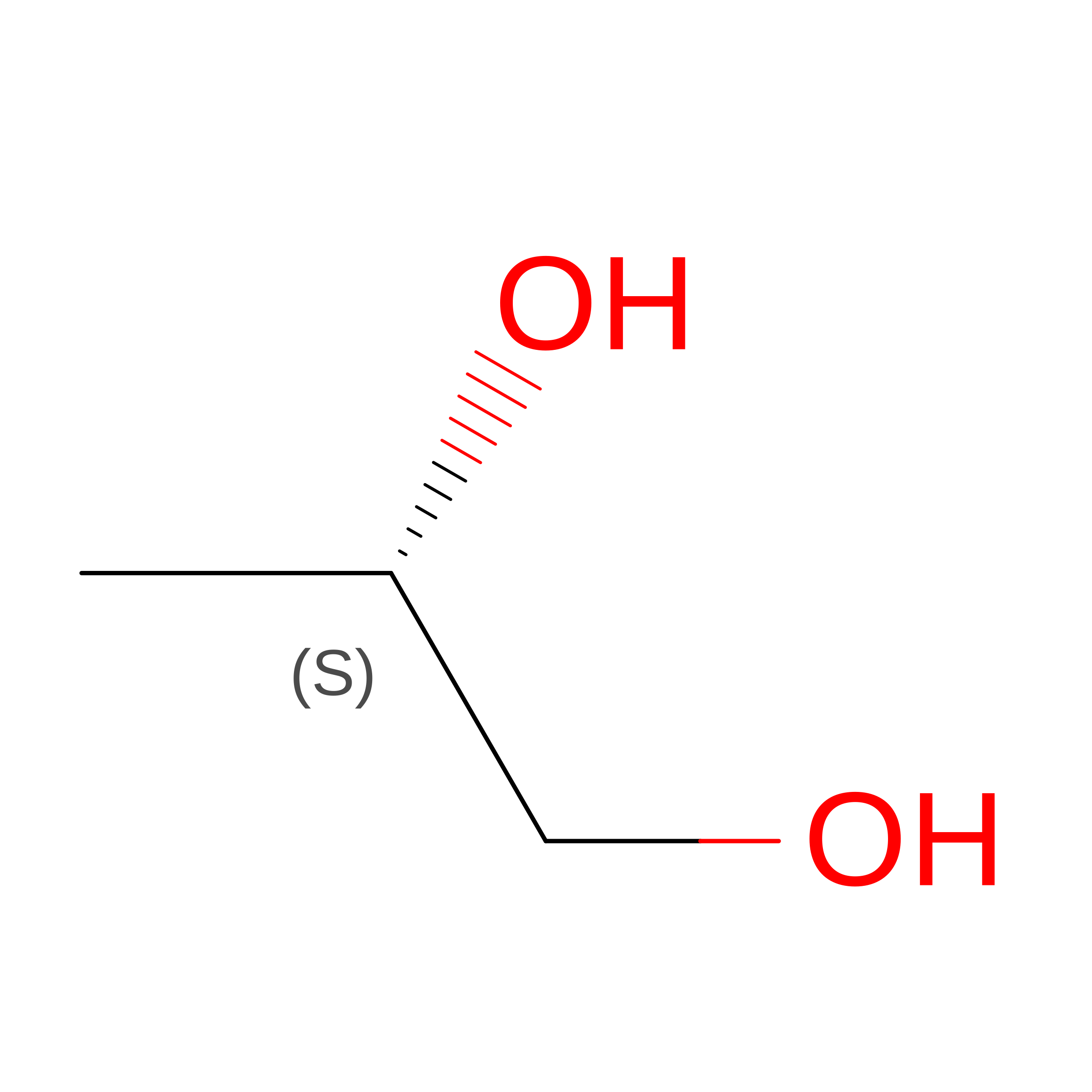
S-1,2-PROPANEDIOL
C3 H8 O2
DNIAPMSPPWPWGF-VKHMYHEASA-N
ZN

Query on ZN


Download:Ideal Coordinates CCD File
D [auth A],
E [auth A],
O [auth B],
P [auth B]
ZINC ION
Zn
PTFCDOFLOPIGGS-UHFFFAOYSA-N
EDO

Query on EDO


Download:Ideal Coordinates CCD File
I [auth A],
T [auth B]
1,2-ETHANEDIOL
C2 H6 O2
LYCAIKOWRPUZTN-UHFFFAOYSA-N
AZI

Query on AZI


Download:Ideal Coordinates CCD File
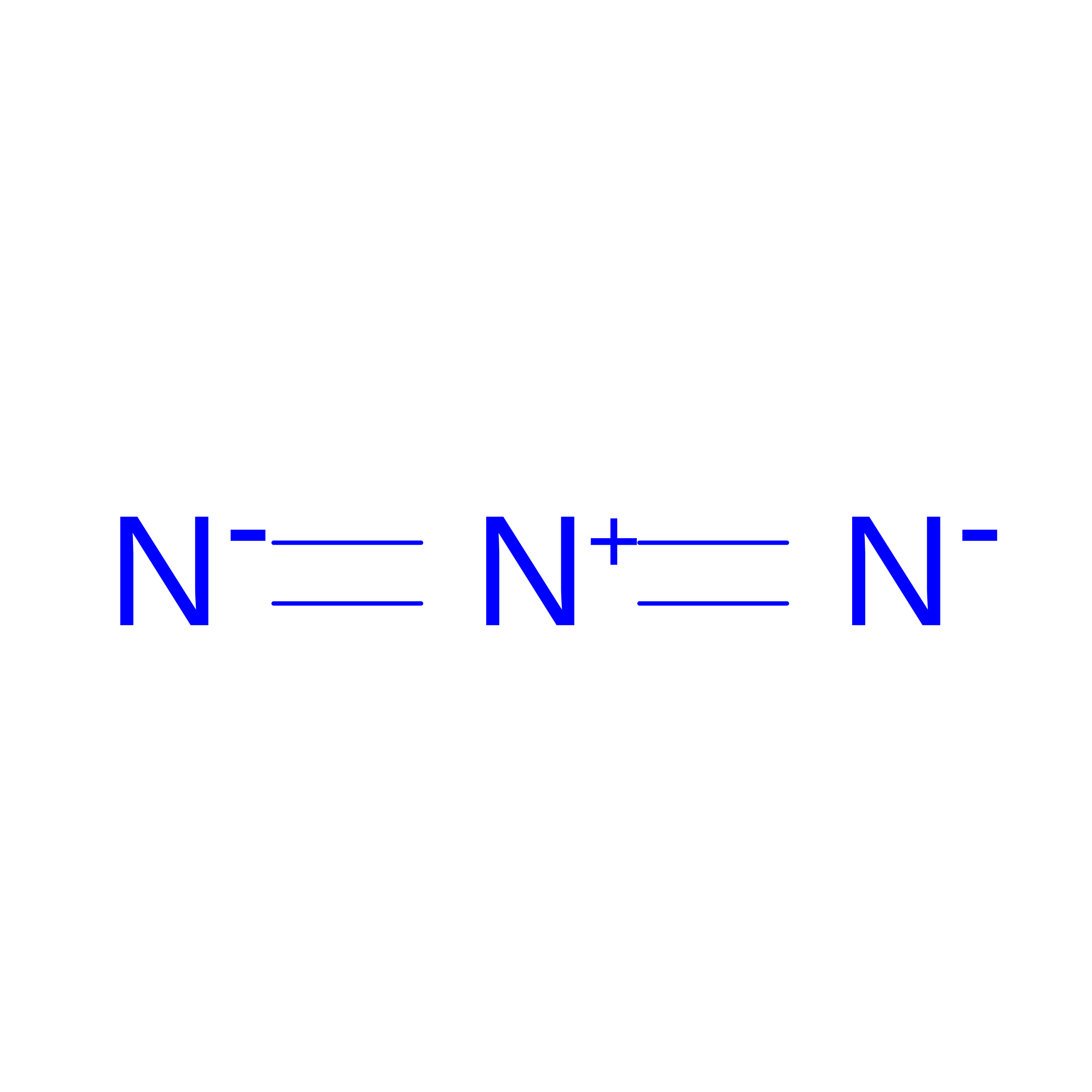
U [auth B]AZIDE ION
N3
IVRMZWNICZWHMI-UHFFFAOYSA-N
CA

Query on CA


Download:Ideal Coordinates CCD File
F [auth A]
G [auth A]
H [auth A]
Q [auth B]
R [auth B]
F [auth A],
G [auth A],
H [auth A],
Q [auth B],
R [auth B],
S [auth B]
CALCIUM ION
Ca
BHPQYMZQTOCNFJ-UHFFFAOYSA-N
NA

Query on NA


Download:Ideal Coordinates CCD File
AA [auth B]
BA [auth B]
V [auth B]
W [auth B]
X [auth B]
AA [auth B],
BA [auth B],
V [auth B],
W [auth B],
X [auth B],
Y [auth B],
Z [auth B]
SODIUM ION
Na
FKNQFGJONOIPTF-UHFFFAOYSA-N

Experimental Data & Validation

Experimental Data

  • Method: X-RAY DIFFRACTION
  • Resolution: 1.65 Å
  • R-Value Free:  0.230 (Depositor), 0.230 (DCC) 
  • R-Value Work:  0.176 (Depositor), 0.177 (DCC) 
  • R-Value Observed: 0.179 (Depositor) 
Space Group: P 1 21 1
Unit Cell:
Length ( Å )Angle ( ˚ )
a = 39.95α = 90
b = 97.39β = 112.72
c = 45.67γ = 90
Software Package:
Software NamePurpose
PHENIXrefinement
XDSdata reduction
XSCALEdata scaling
REFMACphasing

Structure Validation

View Full Validation Report


Ligand Structure Quality Assessment 


Entry History 

Deposition Data

Revision History  (Full details and data files)

  • Version 1.0: 2015-08-26
    Type: Initial release
  • Version 1.1: 2015-10-07
    Changes: Database references
  • Version 1.2: 2024-01-10
    Changes: Data collection, Database references, Derived calculations, Refinement description