4WVL | pdb_00004wvl

Structure-Guided DOT1L Probe Optimization by Label-Free Ligand Displacement


Experimental Data Snapshot

  • Method: X-RAY DIFFRACTION
  • Resolution: 2.41 Å
  • R-Value Free: 
    0.197 (Depositor), 0.200 (DCC) 
  • R-Value Work: 
    0.168 (Depositor), 0.176 (DCC) 
  • R-Value Observed: 
    0.169 (Depositor) 

Starting Model: experimental
View more details

wwPDB Validation 3D Report Full Report

Validation slider image for 4WVL

Ligand Structure Quality Assessment 


This is version 1.4 of the entry. See complete history

Literature

Structure-Guided DOT1L Probe Optimization by Label-Free Ligand Displacement.

Yi, J.S.Federation, A.J.Qi, J.Dhe-Paganon, S.Hadler, M.Xu, X.St Pierre, R.Varca, A.C.Wu, L.Marineau, J.J.Smith, W.B.Souza, A.Chory, E.J.Armstrong, S.A.Bradner, J.E.

(2015) ACS Chem Biol 10: 667-674

  • DOI: https://doi.org/10.1021/cb500796d
  • Primary Citation Related Structures: 
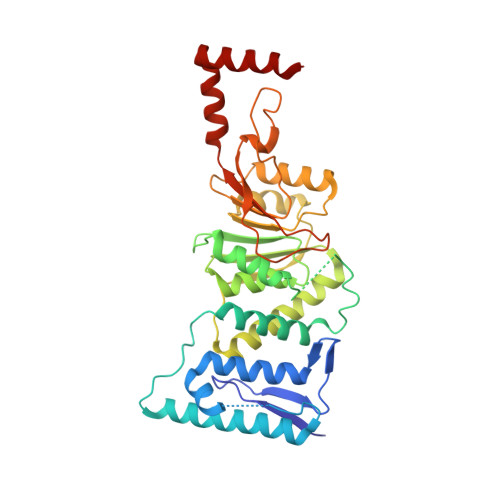
    4WVL

  • PubMed Abstract: 

    The DOT1L lysine methyltransferase has emerged as a validated therapeutic target in MLL-rearranged (MLLr) acute leukemias. Although S-adenosylmethionine competitive inhibitors have demonstrated pharmacological proof-of-principle in MLLr-leukemia, these compounds require further optimization to improve cellular potency and pharmacokinetic stability. Limiting DOT1L inhibitor discovery and ligand optimization have been complex biochemical methods often using radionucleotides and cellular methods requiring prolonged culture. We therefore developed a new suite of assay technologies that allows comparative assessment of chemical tools for DOT1L in a miniaturized format. Coupling these assays with structural information, we developed new insights into DOT1L ligand binding and identified several functionalized probes with increased cellular potency (IC50 values ∼10 nM) and excellent selectivity for DOT1L. Together these assay technologies define a platform capability for discovery and optimization of small-molecule DOT1L inhibitors.


  • Organizational Affiliation
    • †Department of Medical Oncology, Dana-Farber Cancer Institute, Boston, Massachusetts, United States.

Macromolecule Content 

  • Total Structure Weight: 41.54 kDa 
  • Atom Count: 2,779 
  • Modeled Residue Count: 331 
  • Deposited Residue Count: 347 
  • Unique protein chains: 1

Macromolecules

Find similar proteins by:|  3D Structure
Entity ID: 1
MoleculeChains  Sequence LengthOrganismDetailsImage
Histone-lysine N-methyltransferase, H3 lysine-79 specific347Homo sapiensMutation(s): 0 
Gene Names: DOT1LKIAA1814KMT4
EC: 2.1.1.43 (PDB Primary Data), 2.1.1.360 (UniProt)
UniProt & NIH Common Fund Data Resources
Find proteins for Q8TEK3 (Homo sapiens)
Explore Q8TEK3 
Go to UniProtKB:  Q8TEK3
PHAROS:  Q8TEK3
GTEx:  ENSG00000104885 
Entity Groups
Sequence Clusters30% Identity50% Identity70% Identity90% Identity95% Identity100% Identity
UniProt GroupQ8TEK3
Sequence Annotations
Expand
Reference Sequence

Small Molecules

Ligands 4 Unique
IDChains Name / Formula / InChI Key2D Diagram3D Interactions
3US

Query on 3US


Download:Ideal Coordinates CCD File
B [auth A]N-[4-(acetylamino)butyl]-5'-[(3-{[(4-tert-butylphenyl)carbamoyl]amino}propyl)(propan-2-yl)amino]-5'-deoxyadenosine
C33 H51 N9 O5
UMDWALJSYFZDHM-QWOIFIOOSA-N
SO4

Query on SO4


Download:Ideal Coordinates CCD File
C [auth A],
D [auth A],
E [auth A]
SULFATE ION
O4 S
QAOWNCQODCNURD-UHFFFAOYSA-L
GOL

Query on GOL


Download:Ideal Coordinates CCD File
F [auth A]GLYCEROL
C3 H8 O3
PEDCQBHIVMGVHV-UHFFFAOYSA-N
EDO

Query on EDO


Download:Ideal Coordinates CCD File
G [auth A],
H [auth A],
I [auth A]
1,2-ETHANEDIOL
C2 H6 O2
LYCAIKOWRPUZTN-UHFFFAOYSA-N

Experimental Data & Validation

Experimental Data

  • Method: X-RAY DIFFRACTION
  • Resolution: 2.41 Å
  • R-Value Free:  0.197 (Depositor), 0.200 (DCC) 
  • R-Value Work:  0.168 (Depositor), 0.176 (DCC) 
  • R-Value Observed: 0.169 (Depositor) 
Space Group: P 65
Unit Cell:
Length ( Å )Angle ( ˚ )
a = 151.104α = 90
b = 151.104β = 90
c = 53.387γ = 120
Software Package:
Software NamePurpose
PHENIXrefinement
HKL-2000data reduction
HKL-2000data scaling
MOLREPphasing

Structure Validation

View Full Validation Report


Ligand Structure Quality Assessment 


Entry History 

Deposition Data

Revision History  (Full details and data files)

  • Version 1.0: 2014-12-03
    Type: Initial release
  • Version 1.1: 2015-01-28
    Changes: Database references
  • Version 1.2: 2015-04-01
    Changes: Database references
  • Version 1.3: 2015-08-26
    Changes: Data collection
  • Version 1.4: 2023-09-27
    Changes: Data collection, Database references, Derived calculations, Refinement description