4WV7

HEAT SHOCK PROTEIN 70 SUBSTRATE BINDING DOMAIN WITH COVALENTLY LINKED NOVOLACTONE


Experimental Data Snapshot

  • Method: X-RAY DIFFRACTION
  • Resolution: 2.42 Å
  • R-Value Free: 0.272 
  • R-Value Work: 0.200 
  • R-Value Observed: 0.204 

wwPDB Validation   3D Report Full Report


Ligand Structure Quality Assessment 


This is version 1.2 of the entry. See complete history


Literature

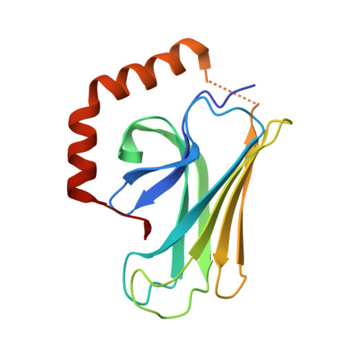
The novolactone natural product disrupts the allosteric regulation of hsp70.

Hassan, A.Q.Kirby, C.A.Zhou, W.Schuhmann, T.Kityk, R.Kipp, D.R.Baird, J.Chen, J.Chen, Y.Chung, F.Hoepfner, D.Movva, N.R.Pagliarini, R.Petersen, F.Quinn, C.Quinn, D.Riedl, R.Schmitt, E.K.Schitter, A.Stams, T.Studer, C.Fortin, P.D.Mayer, M.P.Sadlish, H.

(2015) Chem Biol 22: 87-97

  • DOI: https://doi.org/10.1016/j.chembiol.2014.11.007
  • Primary Citation of Related Structures:  
    4WV5, 4WV7

  • PubMed Abstract: 

    The highly conserved 70 kDa heat shock proteins (Hsp70) play an integral role in proteostasis such that dysregulation has been implicated in numerous diseases. Elucidating the precise role of Hsp70 family members in the cellular context, however, has been hampered by the redundancy and intricate regulation of the chaperone network, and relatively few selective and potent tools. We have characterized a natural product, novolactone, that targets cytosolic and ER-localized isoforms of Hsp70 through a highly conserved covalent interaction at the interface between the substrate-binding and ATPase domains. Biochemical and structural analyses indicate that novolactone disrupts interdomain communication by allosterically inducing a conformational change in the Hsp70 protein to block ATP-induced substrate release and inhibit refolding activities. Thus, novolactone is a valuable tool for exploring the requirements of Hsp70 chaperones in diverse cellular contexts.


  • Organizational Affiliation

    Novartis Institutes for BioMedical Research, 250 Massachusetts Avenue, Cambridge, MA 02139, USA.


Macromolecules
Find similar proteins by:  (by identity cutoff)  |  3D Structure
Entity ID: 1
MoleculeChains Sequence LengthOrganismDetailsImage
Heat shock 70 kDa protein 1A/1B
A, B
151Homo sapiensMutation(s): 0 
Gene Names: HSPA1AHSPA1HSX70HSPA1B
UniProt & NIH Common Fund Data Resources
Find proteins for P0DMV8 (Homo sapiens)
Explore P0DMV8 
Go to UniProtKB:  P0DMV8
PHAROS:  P0DMV8
GTEx:  ENSG00000204389 
Entity Groups  
Sequence Clusters30% Identity50% Identity70% Identity90% Identity95% Identity100% Identity
UniProt GroupP0DMV8
Sequence Annotations
Expand
  • Reference Sequence
Small Molecules
Ligands 1 Unique
IDChains Name / Formula / InChI Key2D Diagram3D Interactions
3UM
Query on 3UM

Download Ideal Coordinates CCD File 
C [auth A],
D [auth B]
(5beta,6alpha,8alpha,14alpha)-13-ethenyl-5,6-dihydroxy-14-methylpodocarp-12-en-15-oic acid
C20 H30 O4
OKUNNUNVWSVFGB-GWXXFZPSSA-N
Experimental Data & Validation

Experimental Data

  • Method: X-RAY DIFFRACTION
  • Resolution: 2.42 Å
  • R-Value Free: 0.272 
  • R-Value Work: 0.200 
  • R-Value Observed: 0.204 
  • Space Group: P 21 21 2
Unit Cell:
Length ( Å )Angle ( ˚ )
a = 67.536α = 90
b = 87.779β = 90
c = 52.453γ = 90
Software Package:
Software NamePurpose
PHASERphasing
BUSTERrefinement
XDSdata reduction
SCALAdata scaling

Structure Validation

View Full Validation Report


Ligand Structure Quality Assessment 


Entry History 

Deposition Data

Revision History  (Full details and data files)

  • Version 1.0: 2015-01-14
    Type: Initial release
  • Version 1.1: 2015-02-04
    Changes: Database references
  • Version 1.2: 2023-09-27
    Changes: Data collection, Database references, Derived calculations, Other, Refinement description, Source and taxonomy