4WUC

N-terminal 43 kDa fragment of the E. coli DNA gyrase B subunit grown from 100 mM NaCl condition


Experimental Data Snapshot

  • Method: X-RAY DIFFRACTION
  • Resolution: 1.90 Å
  • R-Value Free: 0.220 
  • R-Value Work: 0.183 
  • R-Value Observed: 0.185 

wwPDB Validation   3D Report Full Report


This is version 1.2 of the entry. See complete history


Literature

The role of monovalent cations in the ATPase reaction of DNA gyrase

Hearnshaw, S.J.Chung, T.T.Stevenson, C.E.M.Maxwell, A.Lawson, D.M.

(2015) Acta Crystallogr D Biol Crystallogr 71: 996-1005

  • DOI: https://doi.org/10.1107/S1399004715002916
  • Primary Citation of Related Structures:  
    4WUB, 4WUC, 4WUD, 4XTJ

  • PubMed Abstract: 

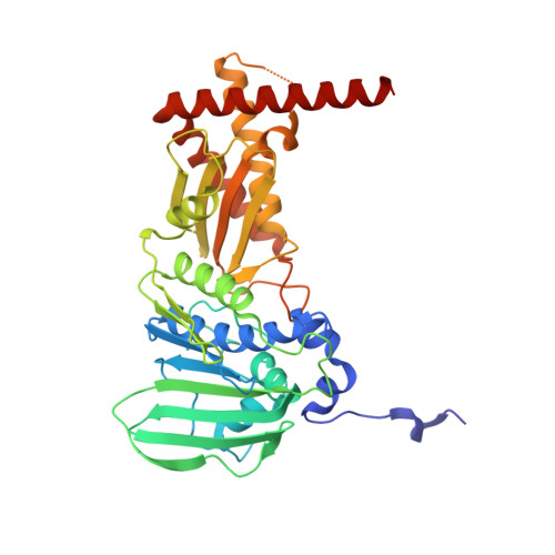
    Four new crystal structures of the ATPase domain of the GyrB subunit of Escherichia coli DNA gyrase have been determined. One of these, solved in the presence of K(+), is the highest resolution structure reported so far for this domain and, in conjunction with the three other structures, reveals new insights into the function of this domain. Evidence is provided for the existence of two monovalent cation-binding sites: site 1, which preferentially binds a K(+) ion that interacts directly with the α-phosphate of ATP, and site 2, which preferentially binds an Na(+) ion and the functional significance of which is not clear. The crystallographic data are corroborated by ATPase data, and the structures are compared with those of homologues to investigate the broader conservation of these sites.


  • Organizational Affiliation

    Department of Biological Chemistry, John Innes Centre, Norwich Research Park, Norwich NR4 7UH, England.


Macromolecules
Find similar proteins by:  (by identity cutoff)  |  3D Structure
Entity ID: 1
MoleculeChains Sequence LengthOrganismDetailsImage
DNA gyrase subunit B392Escherichia coli K-12Mutation(s): 0 
Gene Names: gyrBacrBhimBhisUnalCparApcbAb3699JW5625
EC: 5.99.1.3
UniProt
Find proteins for P0AES6 (Escherichia coli (strain K12))
Explore P0AES6 
Go to UniProtKB:  P0AES6
Entity Groups  
Sequence Clusters30% Identity50% Identity70% Identity90% Identity95% Identity100% Identity
UniProt GroupP0AES6
Sequence Annotations
Expand
  • Reference Sequence
Experimental Data & Validation

Experimental Data

  • Method: X-RAY DIFFRACTION
  • Resolution: 1.90 Å
  • R-Value Free: 0.220 
  • R-Value Work: 0.183 
  • R-Value Observed: 0.185 
  • Space Group: C 2 2 21
Unit Cell:
Length ( Å )Angle ( ˚ )
a = 87.91α = 90
b = 140.88β = 90
c = 79.52γ = 90
Software Package:
Software NamePurpose
REFMACrefinement
Aimlessdata scaling
MOLREPphasing
PDB_EXTRACTdata extraction
XDSdata reduction

Structure Validation

View Full Validation Report


Entry History 

Deposition Data

Revision History  (Full details and data files)

  • Version 1.0: 2015-04-08
    Type: Initial release
  • Version 1.1: 2015-04-22
    Changes: Database references
  • Version 1.2: 2024-01-10
    Changes: Data collection, Database references, Derived calculations, Refinement description